|
Case Report
From mechanism to mitigation: Addressing ponatinib’s cerebral arterial occlusion risk
1 Department of Neurology, Barrow Neurological Institute/Creighton University School of Medicine, Phoenix, Arizona, USA
2 Department of Neurology, Ascension Medical Group, Austin, Texas, USA
Address correspondence to:
Supreet Kaur†
Department of Neurology, University of Texas at Austin/Ascension Medical Group, Austin, Texas,
USA
Message to Corresponding Author
Article ID: 100161Z10MO2026
Access full text article on other devices

Access PDF of article on other devices


How to cite this article
Harnoor V, Kaur S. From mechanism to mitigation: Addressing ponatinib’s cerebral arterial occlusion risk. J Case Rep Images Oncology 2026;12(2):19–25.ABSTRACT
Introduction: Tyrosine kinase inhibitors (TKIs) have significantly improved outcomes in Philadelphia chromosome-positive (Ph+) acute lymphoblastic leukemia (ALL). Ponatinib, a third-generation TKI active against the T315I mutation, offers superior survival benefits but carries an increased risk of arterio-occlusive events (AOEs), including ischemic stroke, particularly in patients with baseline vascular risk factors. We report a suspected ponatinib-associated large vessel occlusion stroke and discuss potential mechanisms, monitoring, and management considerations.
Case Report: A 60-year-old woman with Ph+ ALL on ponatinib for four years, hypertension, diabetes, hyperlipidemia, and prior deep vein thrombosis presented after waking up with dysarthria, left facial weakness, and hemisensory loss. Imaging revealed acute right parietotemporal infarction with proximal right M2 occlusion. She was initially ineligible for intravenous thrombolysis and endovascular thrombectomy (EVT) because of an absent diffusion-weighted imaging–fluid-attenuated inversion recovery mismatch and a low National Institutes of Health Stroke Scale (NIHSS) score. Overnight, she developed dense left hemiplegia and neglect. Statim (STAT) imaging showed favorable perfusion mismatch prompting emergent EVT, which achieved successful reperfusion. Given negative comprehensive embolic stroke workup and right M2 irregularity on follow-up imaging suggestive of intracranial vasculopathy, prolonged ponatinib exposure and uncontrolled vascular risk factors were considered contributory. Ponatinib was discontinued, and she was discharged to rehabilitation with a modified Rankin Scale score of 4 at two months.
Conclusion: This case highlights a suspected ponatinib-associated large vessel ischemic stroke treated with EVT in a patient with prolonged exposure and multiple vascular risk factors. It underscores the risk of cerebrovascular AOEs during ponatinib therapy and importance of vascular risk assessment, monitoring, and multidisciplinary management to reduce treatment-related morbidity while balancing leukemia control.
Keywords: Arterial occlusion, Cerebrovascular disease, Ponatinib, Thrombectomy,
†All work was performed at the Department of Neurology, Barrow Neurological Institute, Phoenix, Arizona.
Introduction
The Philadelphia (Ph) chromosome is pathognomonic for chronic myeloid leukemia (CML) but is also found in 20–30% of adults with acute lymphoblastic leukemia (ALL), where it confers a poorer prognosis [1],[2]. It results from a t(9;22) translocation that creates the BCR-ABL1 fusion gene, which encodes a constitutively active tyrosine kinase, leading to uncontrolled white blood cell proliferation [3].
Targeted therapy with Abl tyrosine kinase inhibitors (TKIs) has dramatically improved outcomes for patients with Ph+ leukemias, increasing five-year overall survival (OS) from 20–50% to over 80%, and enabling long-term survival almost close to that of the general population in many [4].
Imatinib, the first TKI introduced in 2001, offers an OS rate of 43% for Ph+ ALL and 83% for CML, and has an excellent safety profile, notably lacking serious cardiovascular side effects [5]. However, nearly half of patients fail first- and second-generation TKIs (like imatinib and nilotinib, respectively) because of intolerance or resistance resulting from mutations in the BCR-ABL kinase domain [5]. One such first- and second-generation TKI-resistant mutation is the threonine-to-isoleucine mutation at position 315 (T315I) that acts as a “gatekeeper” and prevents TKI binding [5]. It affects up to 75% of relapsed Ph+ ALL patients and is associated with an extremely poor prognosis [6].
Ponatinib, a third-generation TKI, has been shown to be effective against both unmutated and mutated BCR-ABL1 and is the only Food and Drug Administration (FDA)-approved agent for patients with the T315I mutation [7],[8]. Initially FDA-approved for heavily pretreated patients with resistant or progressive disease despite multiple TKI therapies, it is now also considered a frontline option for patients with newly diagnosed Ph+ ALL [5],[7],[8],[9]. However, despite its broad activity and efficacy, ponatinib carries a high risk of serious arterio-occlusive events (AOEs), including stroke and myocardial infarction [7],[10].
The aim of this case report is to highlight the natural course and clinical impact of ponatinib-associated large-vessel occlusion stroke in a patient with vascular risk factors and long-term ponatinib use for Ph+ ALL. We also discuss potential mechanisms, monitoring considerations, and management strategies—including potential endovascular thrombectomy (EVT)—to inform a multidisciplinary approach that may optimize treatment efficacy while reducing disability associated with cerebrovascular adverse events.
Case Report
A 60-year-old woman with a history of hypertension, hyperlipidemia, diabetes, prior unprovoked deep vein thrombosis status post inferior vena cava filter (20 years ago), and Philadelphia chromosome-positive (Ph+) acute lymphoblastic leukemia (ALL; diagnosed four years ago) presented after waking up with acute-onset headache, left facial droop, dysarthria, and left-sided paresthesias. Cerebral imaging (Figure 1A, Figure 1B, Figure 1C) demonstrated acute right parietotemporal infarcts and proximal right M2 segment occlusion. However, the patient was ineligible for intravenous thrombolysis or EVT. Intravenous thrombolysis was not indicated because the patient did not meet the WAKE-UP trial criteria [11], presenting 6 hours after awakening and lacking a diffusion-weighted imaging–fluid-attenuated inversion recovery (DWI–FLAIR) mismatch. Endovascular thrombectomy was not pursued because of a low NIHSS score of 3.
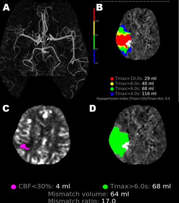
She was admitted in stable condition but experienced early neurological deterioration (END) 12 hours later, developing acute left-sided paralysis and hemineglect. STAT imaging (Figure 2A, Figure 2B, Figure 2C, Figure 2D) confirmed persistent right M2 occlusion, with favorable perfusion profile [penumbra/tissue-at-risk defined as Tmax > 6 seconds of 68 mL, ischemic core defined as cerebral blood flow (CBF) of <30% of 4 mL, mismatch/salvageable tissue volume of 64 mL]. Consequently, she underwent emergent EVT with thrombolysis in cerebral infarction 2c reperfusion after three passes (Figure 3A, Figure 3B, Figure 3C, Figure 3D). No overt underlying intracranial disease was noted post-recanalization. Follow-up imaging (Figure 1D, Figure 1E, Figure 1F) showed significant progression of the right parietotemporal infarction.
Standard stroke workup including computed tomography angiography showed no significant extracranial or intracranial large artery atherosclerotic disease or non-stenotic carotid plaque. Echocardiogram showed normal ejection fraction, atrium size, and valve function, no regional wall motion abnormality or patent foramen ovale, and a normal aortic root. Inpatient telemetry and outpatient 14-day Holter monitoring showed normal sinus rhythm. Vascular risk factors were noted to be uncontrolled: LDL was elevated at 114 mg/dL and hemoglobin A1c at 8.8%; however, these risk factors in isolation did not explain her large vessel occlusive event.
Medication review revealed daily ponatinib use. The patient reported that she was diagnosed with Ph+ ALL four years prior. Due to chemotherapy intolerance, she was initiated on ponatinib therapy at that time. She reported being in remission and taking ponatinib at a dose of 15 mg daily with prophylactic apixaban to mitigate the AOE risk associated with ponatinib. However, incomplete adherence to apixaban was noted as the patient was taking it once daily.
Based on this history and given large vessel occlusion without clear embolic source on standard stroke workup, cancer-associated hypercoagulability, and current tyrosine kinase inhibitor (TKI)-related vasculopathy were considered. Hematology confirmed minimal residual disease negativity (no detectable cancer cells on reverse transcriptase–quantitative polymerase chain reaction). A hematology-directed hypercoagulable panel performed following apixaban discontinuation was unrevealing, with negative antiphospholipid antibodies (anticardiolipin IgG/IgM/IgA and β2-glycoprotein IgG/IgM < 10, no confirmatory evidence of lupus anticoagulant), and negative testing for Factor V Leiden and prothrombin G20210A mutations.
Given the absence of an alternative stroke mechanism and the patient’s prolonged exposure to ponatinib, a ponatinib-associated cerebrovascular AOE was considered the most likely contributor to her stroke. Ponatinib was subsequently discontinued, and apixaban was not deemed necessary for secondary prophylaxis by the patient’s hematologist. The patient was discharged to inpatient rehabilitation on aspirin. At a two-month follow-up, her modified Rankin Scale score was 4 (representing Moderately-severe disability marked by inability to ambulate independently and needing help with activities of daily living) and follow-up imaging showed irregularity of right M2 (Figure 3E and Figure 3F).


Discussion
Ponatinib, a third-generation TKI, is the only FDA-approved agent for Ph+ leukemia with the T315I mutation, which confers resistance to earlier-generation TKIs and is associated with a poor prognosis [7],[8]. Its accelerated approval in 2012 was based on the landmark single-arm, phase II Ponatinib Ph-positive ALL and CML Evaluation (PACE) trial, which, while highlighting its broad antileukemic activity, also revealed a substantial risk of serious arterio-occlusive events (AOEs; 25% non-adjudicated, 17% adjudicated), including stroke and myocardial infarction [5]. These serious adverse events typically occurred later during therapy (median time to stroke, 1.4 years), were more common at higher doses (with each 15-mg dose reduction predicting an approximately 33% reduction in AOE risk), and occurred more frequently in patients with pre-existing vascular risk factors, although they were also reported in younger patients without cardiovascular comorbidities [5],[10].
These findings prompted the Optimizing Ponatinib Treatment in CP-CML (OPTIC) trial, which showed that response-based dose reduction—i.e., starting at 45 mg until response, then decreasing to 15 mg—significantly lowers AOEs compared with historical fixed-dose strategies [12]. The trial also demonstrated that older age (≥65 years) was independently associated with higher AOE rates (38% vs 8%) and increased exposure correlated with AOE risk; however, exposure-adjusted event rates decreased over time [12].
Similarly, the Ponatinib vs Imatinib in Frontline Ph-positive ALL trial (PhALLCON) applied a response-based dosing strategy (starting at 30 mg until response, then reducing to 15 mg), excluded patients with significant vascular risk factors, and reported comparable and infrequent AOEs [9]. The authors attributed the low AOE rate to a “reduced starting dose, proactive dose reduction, stringent exclusion criteria, and a less heavily pretreated population” [9].
Despite demonstrating that response-based dosing can reduce AOE risk, both OPTIC and PhALLCON excluded patients with significant vascular risk factors, limiting the generalizability of their safety findings [9],[12]. This limitation is particularly noteworthy because analysis of a real-world claims database by Jabbour et al. found that more than 60% of patients with CML had pre-existing vascular risk factors at the time of TKI initiation [13]. Furthermore, ponatinib itself is associated with hypertension [7],[10].
Accordingly, important questions remain regarding the underlying mechanisms of ponatinib-associated cerebrovascular AOEs, particularly given that treatment duration may range from a minimum of two years to lifelong therapy, and that stroke can severely impact quality of life [14].
To address this knowledge gap, we synthesized key findings regarding ponatinib-associated cerebrovascular toxicity. Current evidence links ponatinib-associated cerebrovascular AOEs to intracranial stenosis, predominantly affecting large arteries [15],[16] [17],[18],[19]. Whether ponatinib causes or exacerbates this vasculopathy remains debated; however, its prevalence in patients with vascular risk factors suggests the latter. As AOEs occur more frequently than venous thromboembolism, platelet-and atherosclerotic plaque-mediated mechanisms are likely involved. This is substantiated by murine model experiments demonstrating that ponatinib triggers plaque inflammation and spontaneous plaque rupture by activating platelets, leukocytes, and endothelial cell TNF (tumor necrosis factor) signaling [20],[21],[22]. Via Notch-1 hyperactivation, ponatinib reduces endothelial survival and enhances senescence and apoptosis, while heightened arterial thrombosis and platelet reactivity drastically reduce carotid occlusion time (from ~32 to 10 minutes) [20],[21],[22]. Notably, these effects are attenuated in murine models by selective Notch-1 blockade, tumor necrosis factor receptor (TNFR)-inhibition, or pioglitazone [20],[21],[22]. Overall, these findings give credence to the possibility that ponatinib-associated stroke likely occurs due to accelerated intracranial atherosclerotic vasculopathy.
Applying this hypothesis to our patient, we sought to determine if her occlusion was consistent with intracranial atherosclerosis-related large vessel occlusion (ICAD-LVO), using established clinical and imaging/procedural criteria [23],[24]. Features suggestive of ICAD-LVO include fluctuating neurological deficits, absence of atrial fibrillation, truncal-type occlusion with tapered narrowing proximal to the occlusion, and residual stenosis with early reocclusion post-EVT [23],[24].
Our patient exhibited several ICAD-LVO characteristics, including fluctuating symptoms with mild deficits at onset followed by early neurological deterioration, a relatively small infarct core on perfusion imaging, and intracranial atherosclerotic disease in other vascular territories (bilateral posterior cerebral arteries). However, she lacked classic features such as extra- or intracranial arterial calcification, distal hypoperfusion or watershed infarction (reflecting hemodynamic compromise from pre-existing stenosis), tapered narrowing proximal to the occlusion, residual fixed stenosis >50% after thrombectomy, and early reocclusion. While this raises the possibility of a cryptogenic mechanism for her stroke, the combination of uncontrolled vascular risk factors, prolonged ponatinib exposure, and persistent vessel irregularity on follow-up imaging two months later support an underlying intracranial vasculopathy. Notably, to our knowledge, this case provides unique longitudinal pre- and post-thrombectomy vascular imaging in suspected ponatinib-related large vessel occlusion (Figure 3A, Figure 3B, Figure 3C, Figure 3D, Figure 3E, Figure 3F).
Taken together, these findings suggest that cerebrovascular AOEs may be mitigated—not only through response-based dose optimization, as highlighted in OPTIC and PhALLCON, but also through comprehensive cardiovascular risk assessment, aggressive risk factor stratification and serial vascular imaging in selected high-risk patients.
Risk stratification tools may help identify patients at increased risk of cardiovascular events during ponatinib therapy. The German Consensus Recommendations suggest applying general cardiovascular guidelines such as the European Society of Cardiology’s SCORE2 (age < 70) and SCORE2-OP (age > 70) [25]. Consistent with this approach, a retrospective study of 85 patients with Ph+ leukemias found these scores predictive of cardiovascular risk and potentially useful for patient attention and TKI selection [26]. Additionally, the Campus CML study linked lower plasma LDL (<70 mg/dL), cholesterol, and triglycerides with reduced AOEs in real-world ponatinib-treated CML patients [27].
Beyond risk factor control, surveillance may enable earlier detection of cerebrovascular complications. Because intracranial stenosis may progress silently, mandatory cerebral imaging—preferably magnetic resonance angiography or computed tomography angiography—should be done at baseline and during TKI therapy. Notably, 95% of adjudicated AOEs in the PACE were serious despite antiplatelet use, and although overall survival was not adversely affected, stroke can substantially impair quality of life as demonstrated by the clinical impact on our patient [10]. These considerations highlight the importance of a stroke neurologist as part of the multidisciplinary team, alongside the emerging field of cardio-oncology. Given ponatinib’s cardiac side-effects (cardiac failure, atrial fibrillation, myocardial infarction), patients should undergo periodic echocardiograms, and, if indicated, cardiac monitoring for prevention of cardioembolic stroke [7].
The role of pharmacological prophylaxis, including antithrombotic therapy, remains uncertain. In PACE, which enrolled patients under less restrictive vascular risk criteria, AOEs occurred despite relatively high rates of antiplatelet use; 47% (37/78) of patients with AOEs had received concomitant platelet aggregation inhibitors at some point during treatment [10]. In contrast, a retrospective study of 85 adult patients with CML treated with ponatinib suggested a lower AOE incidence in patients =60 years treated with aspirin compared with those not receiving aspirin (33.3% vs 61.8%) [26]. Other preventative strategies like pioglitazone, selective Notch-1 blockers, and TNFR-mediated endothelial activation blockers also warrant further investigation [20],[21],[22].
Although preventive strategies remain an active area of investigation, management after stroke onset also presents unique challenges. Despite initially low neurological severity, most cases report a pattern of END. Previous authors used superficial temporal artery-middle cerebral artery (STA-MCA) bypass for END in patients with long-segment disease who deteriorated late (3–7 days), precluding endovascular revascularization [15],[16],[17],[18]. However, we performed EVT due to the patient’s timely presentation (<24 hours) and achieved successful reperfusion without any clinically significant complications. Evaluation of clot composition was not performed, but it would be helpful to investigate in the future to better elucidate the pathophysiology of ponatinib associated cerebrovascular AOEs.
Conclusion
Ponatinib-associated cerebrovascular AOEs represent an infrequent but potentially devastating complication of an otherwise highly efficacious therapy for Ph+ ALL. Our case illustrates that prolonged ponatinib use, particularly in the presence of uncontrolled vascular risk factors, may contribute to intracranial vasculopathy and a highly disabling large vessel occlusion stroke. While this report suggests a potential link, it is critical to emphasize that a definitive causal relationship cannot be established from a single instance; these findings should be interpreted as an association rather than conclusive evidence of causation. Clinically, this case illustrates that patients may present with mild initial deficits followed by early neurological deterioration, underscoring the critical need for vigilant monitoring and the rapid identification of candidates for acute interventions like EVT. Ultimately, this report reinforces the necessity of multidisciplinary collaboration between hematologists, vascular neurologists, and cardiologists to optimize cerebrovascular safety while maintaining leukemia control. Future research should focus on elucidating the specific mechanisms underlying cerebrovascular AOEs, improving risk stratification and vascular surveillance protocols, and investigating targeted preventive strategies to reduce treatment-related morbidity while preserving the therapeutic benefits of ponatinib.
REFERENCES
1.
Burmeister T, Schwartz S, Bartram CR, Gökbuget N, Hoelzer D, Thiel E, et al. Patients’ age and BCR-ABL frequency in adult B-precursor ALL: A retrospective analysis from the GMALL study group. Blood 2008;112(3):918–9. [CrossRef]
[Pubmed]

2.
Wetzler M, Dodge RK, Mrózek K, Carroll AJ, Tantravahi R, Block AW, et al. Prospective karyotype analysis in adult acute lymphoblastic leukemia: The cancer and leukemia Group B experience. Blood 1999;93(11):3983–93.
[Pubmed]
3.
Rowley JD. Letter: A new consistent chromosomal abnormality in chronic myelogenous leukaemia identified by quinacrine fluorescence and Giemsa staining. Nature 1973;243(5405):290–3. [CrossRef]
[Pubmed]
4.
Sasaki K, Haddad FG, Short NJ, Jain N, Issa G, Jabbour E, et al. Outcome of Philadelphia chromosomepositive chronic myeloid leukemia in the United States since the introduction of imatinib therapy—The Surveillance, Epidemiology, and End Results database, 2000–2019. Cancer 2023;129(23):3805–14. [CrossRef]
[Pubmed]
5.
Cortes JE, Kim DW, Pinilla-Ibarz J, le Coutre P, Paquette R, Chuah C, et al. A phase 2 trial of ponatinib in Philadelphia chromosome-positive leukemias. N Engl J Med 2013;369(19):1783–96. [CrossRef]
[Pubmed]
6.
Rousselot P, Coudé MM, Gokbuget N, Gambacorti Passerini C, Hayette S, Cayuela JM, et al. Dasatinib and low-intensity chemotherapy in elderly patients with Philadelphia chromosome-positive ALL. Blood 2016;128(6):774–82. [CrossRef]
[Pubmed]
7.
Pulte ED, Chen H, Price LSL, Gudi R, Li H, Okusanya OO, et al. FDA Approval summary: Revised indication and dosing regimen for ponatinib based on the results of the OPTIC trial. Oncologist 2022;27(2):149–57. [CrossRef]
[Pubmed]
8.
Shah B, Mattison RJ, Abboud R, Abdelmessieh P, Aldoss I, Burke PW, et al. Acute lymphoblastic leukemia, version 2.2024, NCCN Clinical Practice Guidelines in Oncology. J Natl Compr Canc Netw 2024;22(8):563–76. [CrossRef]
[Pubmed]
9.
Jabbour E, Kantarjian HM, Aldoss I, Montesinos P, Leonard JT, Gómez-Almaguer D, et al. Ponatinib vs imatinib in frontline Philadelphia chromosome-positive acute lymphoblastic leukemia: A randomized clinical trial. JAMA 2024;331(21):1814–23. [CrossRef]
[Pubmed]
10.
Januzzi JL, Garasic JM, Kasner SE, McDonald V, Petrie MC, Seltzer J, et al. Retrospective analysis of arterial occlusive events in the PACE trial by an independent adjudication committee. J Hematol Oncol 2022;15(1):1. [CrossRef]
[Pubmed]
11.
Thomalla G, Simonsen CZ, Boutitie F, Andersen G, Berthezene Y, Cheng B, et al. MRI-guided thrombolysis for stroke with unknown time of onset. N Engl J Med 2018;379(7):611–22. [CrossRef]
[Pubmed]
12.
Cortes J, Apperley J, Lomaia E, Moiraghi B, Undurraga Sutton M, Pavlovsky C, et al. Ponatinib dose-ranging study in chronic-phase chronic myeloid leukemia: A randomized, open-label phase 2 clinical trial. Blood 2021;138(21):2042–50. [CrossRef]
[Pubmed]
13.
Jabbour E, Makenbaeva D, Lingohr-Smith M, Lin J. Use of real-world claim databases to assess prevalence of comorbid conditions relevant to the treatment of chronic myelogenous leukemia based on National Comprehensive Network Treatment Guidelines. Clin Lymphoma Myeloma Leuk 2015;15(12):797–802. [CrossRef]
[Pubmed]
14.
Kugler E, Kantarjian H, Jabbour E, Khaire N, Short NJ, Kadia TM, et al. Treatment-free remission in nontransplanted patients with Philadelphia chromosome-positive acute lymphoblastic leukemia. Cancer 2025;131(5):e35773. [CrossRef]
[Pubmed]
15.
Spina E, Renna R, Lanterna LA, Colleoni ML, Andreone V. Progressive thrombosis of cervical and intracranial arteries related to Ponatinib treatment for chronic myeloid leukemia. J Stroke Cerebrovasc Dis 2020;29(9):105085. [CrossRef]
[Pubmed]
16.
Azad F, Zhang J, Miranda CJ, Gravina M. Ponatinibinduced cerebrovascular accident (CVA). Cureus 2022;14(12):e32383. [CrossRef]
[Pubmed]
17.
Hirayama A, Sorimachi T, Yokota K, Shigematsu H, Srivatanakul K, Matsumae M. New-generation tyrosine kinase inhibitor-associated asymptomatic cerebrovascular stenosis: Two illustrative cases. Acta Neurochir (Wien) 2022;164(6):1623–6. [CrossRef]
[Pubmed]
18.
Hanazono A, Abe M, Togashi S, Takahashi T, Takahashi N, Sugawara M. Case report: Three characteristics of tyrosine kinase inhibitor-associated cerebrovascular stenosis. High threshold for infarction, atypical infarct area, and vascular recoverability under the use of ponatinib. Front Stroke 2024;3:1367869. [CrossRef]
[Pubmed]
19.
Sasaki N, Enomoto Y, Hori T, Matsubara H, Egashira Y, Izumo T. Cerebrovascular stenosis related to tyrosine kinase inhibitor for chronic myeloid leukemia: Two illustrative cases. BMC Neurol 2025;25(1):6. [CrossRef]
[Pubmed]
20.
Stepanian A, Travers RJ, Tesfu A, Wolter NL, Man JJ, Lu Q, et al. Ponatinib, but not the new Abl-kinase inhibitor asciminib, activates platelets, leukocytes, and endothelial cell TNF signaling to induce atherosclerotic plaque inflammation, myocardial infarction, and stroke. Circulation 2025;152(11):765–83. [CrossRef]
[Pubmed]
21.
Madonna R, Pieragostino D, Cufaro MC, Doria V, Del Boccio P, Deidda M, et al. Ponatinib induces vascular toxicity through the Notch-1 signaling pathway. J Clin Med 2020;9(3):820. [CrossRef]
[Pubmed]
22.
Merkulova A, Mitchell SC, Stavrou EX, Forbes GL, Schmaier AH. Ponatinib treatment promotes arterial thrombosis and hyperactive platelets. Blood Adv 2019;3(15):2312–6. [CrossRef]
[Pubmed]
23.
de Havenon A, Zaidat OO, Amin-Hanjani S, Nguyen TN, Bangad A, Abbasi M, et al. Large vessel occlusion stroke due to intracranial atherosclerotic disease: Identification, medical and interventional treatment, and outcomes. Stroke 2023;54(6):1695–705. [CrossRef]
[Pubmed]
24.
Psychogios M, Brehm A, López-Cancio E, Marco De Marchis G, Meseguer E, Katsanos AH, et al. European Stroke Organisation guidelines on treatment of patients with intracranial atherosclerotic disease. Eur Stroke J 2022;7(3):III–IV. [CrossRef]
[Pubmed]
25.
Saussele S, La Rosée P, Kiani A, Haverkamp W, Jentsch-Ullrich K, Stegelmann F, et al. Addendum to the German consensus recommendations on ponatinib in the treatment of chronic myeloid leukemia. Acta Haematol 2024;147(3):344–51. [CrossRef]
[Pubmed]
26.
Mulas O, Abruzzese E, Luciano L, Iurlo A, Attolico I, Castagnetti F, et al. The new Systematic Coronary Risk Evaluation (SCORE2 and SCORE2-OP) estimates the risk of arterial occlusive events in chronic myeloid leukemia patients treated with nilotinib or ponatinib. Ann Hematol 2024;103(2):427–36. [CrossRef]
[Pubmed]
27.
Caocci G, Mulas O, Capodanno I, Abruzzese E, Iurlo A, Luciano L, et al. Low low-density lipoprotein (LDL), cholesterol and triglycerides plasma levels are associated with reduced risk of arterial occlusive events in chronic myeloid leukemia patients treated with ponatinib in the real-life. A Campus CML study. Blood Cancer J 2020;10(6):66. [CrossRef]
[Pubmed]
SUPPORTING INFORMATION
Acknowledgments
We acknowledge the patient and the excellent clinical team.
Artificial intelligence (AI) use by authors
Online free version of ChatGPT was used for spelling, thesaurus, and grammar checks.
Vishal Harnoor† - Acquisition of data, Analysis of data, Drafting the work, Final approval of the version to be published, Agree to be accountable for all aspects of the work in ensuring that questions related to the accuracy or integrity of any part of the work are appropriately investigated and resolved.
Supreet Kaur† - Conception of the work, Design of the work, Acquisition of data, Analysis of data, Drafting the work, Revising the work critically for important intellectual content, Final approval of the version to be published, Agree to be accountable for all aspects of the work in ensuring that questions related to the accuracy or integrity of any part of the work are appropriately investigated and resolved.
Guaranter of SubmissionThe corresponding author is the guarantor of submission.
Source of SupportNone
Consent StatementWritten informed consent was obtained from the patient for publication of this article.
Data AvailabilityAll relevant data are within the paper and its Supporting Information files.
Conflict of InterestAuthors declare no conflict of interest.
Copyright© 2026 Vishal Harnoor et al. This article is distributed under the terms of Creative Commons Attribution License which permits unrestricted use, distribution and reproduction in any medium provided the original author(s) and original publisher are properly credited. Please see the copyright policy on the journal website for more information.