|
Clinical Image
Metastatic Merkel cell carcinoma: Pancreatic mass, an unknown primary site
1 MD, Department of Internal Medicine, St Luke’s University Health Network, Bethlehem, PA, USA
2 MD, Medical Director of Genitourinary Cancer Program, St Luke’s University Health Network, Bethlehem, PA, USA
3 MD, Medical Director, Clinical Flow Cytometry, St Luke’s University Health Network, Bethlehem, PA, USA
Address correspondence to:
Aashima Gupta
MD, 801 Ostrum Street, Bethlehem, PA,
USA
Message to Corresponding Author
Article ID: 100157Z10AG2026
Access full text article on other devices

Access PDF of article on other devices


How to cite this article
Gupta A, Arriaga Y, Plowman R. Metastatic Merkel cell carcinoma: Pancreatic mass, an unknown primary site. J Case Rep Images Oncology 2026;12(2):1–3.ABSTRACT
No Abstract
Keywords: Merkel cell carcinoma, Pancreatic malignancy, Unknown primary malignancy
Case Report
A 73-year-old man with known history of paroxysmal atrial fibrillation, stage III chronic kidney disease, and hypertriglyceridemia presented with a one-month history of progressive epigastric pain, shortness of breath, nausea, loss of appetite, and unintentional weight loss of approximately 40 pounds. His family noticed pallor paler and jaundiced. The patient also noted dark yellow urine. Upon arrival, vitals were stable with blood pressure (BP) 117/71 mmHg, heart rate 74 beats/minute, temperature was normal (afebrile), oxygen saturation 97% on room air. Initial blood work with unremarkable complete blood count (CBC) with White cell count (WBC) 5.03 × 10³/µL (reference: 4–10 × 103/µL), hemoglobin (Hgb) 15.0 g/dL (reference: 13–16 g/dL), and platelets 280 × 103/µL (reference: 149–390 × 103/µL). Comprehensive metabolic panel (CMP) was abnormal with elevated creatinine 2.13 mg/dL (Baseline creatinine 0.9–1.1 mg/dL), Blood urea nitrogen (BUN) 56 mg/dL (5–25 mg/dL), elevated liver enzymes: aspartate aminotransferase (AST) 90 U/L (reference: 13–39 U/L), alanine aminotransferase (ALT) 155 U/L (7–52 U/L), alkaline phosphatase 288 U/L (34–104 U/L), and total bilirubin 11.06 mg/dL (0.20–1.00 mg/dL). Lipase was also elevated to 2551 U/L (11–88 U/L). Urine studies with some ketonuria no proteinuria, hematuria, pyuria, or bilirubinuria noted.
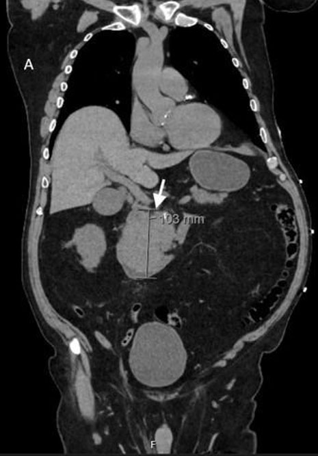
Contrast-enhanced computed tomography (CECT) of the abdomen and pelvis performed during initial workup revealed a large lobulated mass arising from the pancreatic head and uncinate process, measuring 10.2 × 7.9 × 10.3 cm, with associated biliary ductal dilatation, highly suspicious for pancreatic malignancy (Figure 1). Given high-risk features including unintentional 40 pound weight loss over one month, initial imaging suggestive of pancreatic mass, the patient was admitted for further evaluation of possible pancreatic malignancy. This was followed by magnetic resonance cholangiopancreatography (MRCP), which confirmed an approximately 10 cm pancreatic head mass encasing the proximal superior mesenteric artery and occluding the superior mesenteric vein, with no evidence of distant metastasis.
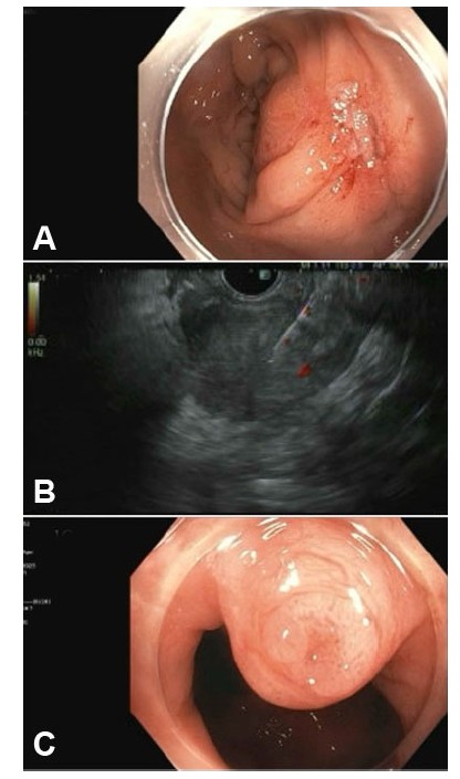
Endoscopic retrograde cholangiopancreatography (ERCP) demonstrated an ulcerated mass within the duodenum. The common bile duct was markedly dilated up to 19 mm, with an abrupt distal cutoff and replacement of the ampulla by tumor. Endoscopic ultrasound identified a heterogeneous, hypoechoic, irregular solid mass measuring 76 × 89 mm within the pancreatic head. Endoscopic ultrasonography-guided biliary stent placement and biopsy of the mass were performed.
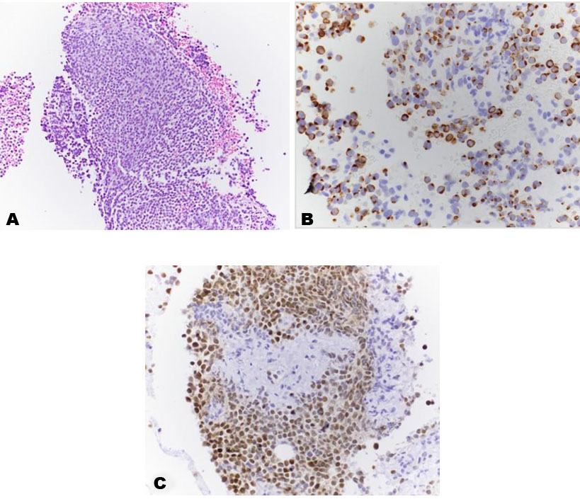
Core-needle biopsy showed sheets of monotonous, small- to medium-sized malignant cells with finely stippled chromatin and inconspicuous nucleoli (Figure 2A, hematoxylin and eosin stain). Initial diagnostic consideration included neuro-endocrine tumor and lymphoma. Subsequently, flow cytometry demonstrated malignant cells were CD56 positive and CD 45 negative suggestive of non-hematolymphoid origin. Due to concern of neuro-endocrine nature immunohistochemistry was performed which demonstrated positivity for CK20 in a perinuclear dot-like pattern (Figure 2B), along with CAM 5.2 and chromogranin A. The Ki-67 proliferative index exceeded 90%, consistent with a high-grade neuroendocrine carcinoma. Tissue was sent for next-generation sequencing (NGS), which identified a tumor profile most compatible with Merkel cell carcinoma. This was further supported by a positive Merkel cell polyomavirus (MCPyV) immunostain (Figure 2C), confirming metastatic Merkel cell carcinoma from an unknown primary source.
The patient was started on first-line systemic chemotherapy with modified FOLFIRINOX (infusional 5-fluorouracil, oxaliplatin, and irinotecan) and achieved a near-complete radiographic response. Dermatology was consulted to evaluate for a primary cutaneous lesion; however, no primary skin lesion was identified.
Discussion
Merkel cell carcinoma (MCC) is a rare and aggressive cutaneous neuroendocrine malignancy with an estimated annual incidence of approximately 2448 cases worldwide [1]. It most commonly spreads to regional lymph nodes, followed by bone and bone marrow, lung, and skin [2]. Pancreatic metastasis is exceedingly rare.
When MCC does metastasize to the pancreas, it can closely mimic a primary pancreatic neuroendocrine neoplasm, creating significant diagnostic difficulty. The distinction is important because the two conditions are managed very differently and carry very different prognoses.
In our patient, next gene sequencing (NGS) played a central role in establishing the correct diagnosis. It identified the pancreatic lesion as metastatic MCC in the absence of any detectable primary tumor, which was further supported by positive MCPyV immunostaining. This finding substantially altered both disease staging and treatment planning. While localized pancreatic neuroendocrine tumors are typically managed with surgical resection [3]. Metastatic MCC is better approached with systemic therapy [4],[5]. Immune checkpoint inhibitors targeting the PD-1 pathway have shown meaningful efficacy in metastatic MCC [4],[5],[6]. Our patient’s plan includes transition to maintenance immunotherapy following his chemotherapy response, with the goal of achieving durable disease control.
Conclusion
This case represents a rare presentation of metastatic Merkel cell carcinoma involving the pancreas with no identifiable primary skin lesion. It highlights two important points: first, MCC should be kept in mind when evaluating pancreatic masses, even in the absence of a known cutaneous primary; and second, next-generation sequencing can be a valuable and at times essential diagnostic tool in atypical cases. In this patient, NGS not only confirmed the diagnosis but also changed the staging and guided appropriate systemic treatment.
REFERENCES
1.
Paulson KG, Park SY, Vandeven NA, Lachance K, Thomas H, Chapuis AG, et al. Merkel cell carcinoma: Current US incidence and projected increases based on changing demographics. J Am Acad Dermatol 2018;78(3):457–63.e2. [CrossRef]
[Pubmed]

2.
Akaike G, Akaike T, Fadl SA, Lachance K, Nghiem P, Behnia F. Imaging of Merkel cell carcinoma: What imaging experts should know. Radiographics 2019;39(7):2069–84. [CrossRef]
[Pubmed]
3.
Akirov A, Larouche V, Alshehri S, Asa SL, Ezzat S. Treatment options for pancreatic neuroendocrine tumors. Cancers (Basel) 2019;11(6):828. [CrossRef]
[Pubmed]
4.
Park SY, Nghiem PT, Tai P, Silk AW. Treatment of recurrent and metastatic Merkel cell carcinoma. UpToDate 2026;11.0.
5.
Gauci ML, Aristei C, Becker JC, Blom A, Bataille V, Dreno B, et al. Diagnosis and treatment of Merkel cell carcinoma: European consensus-based interdisciplinary guideline – Update 2022. Eur J Cancer 2022;171:203–31. [CrossRef]
[Pubmed]
6.
Cassler NM, Merrill D, Bichakjian CK, Brownell I. Merkel cell carcinoma therapeutic update. Curr Treat Options Oncol 2016;17(7):36. [CrossRef]
[Pubmed]
SUPPORTING INFORMATION
Author Contributions
Aashima Gupta - Conception of the work, Design of the work, Acquisition of data, Drafting the work, Final approval of the version to be published, Agree to be accountable for all aspects of the work in ensuring that questions related to the accuracy or integrity of any part of the work are appropriately investigated and resolved.
Yull Arriaga - Drafting the work, Final approval of the version to be published, Agree to be accountable for all aspects of the work in ensuring that questions related to the accuracy or integrity of any part of the work are appropriately investigated and resolved.
Russell Plowman - Acquisition of data, Revising the work critically for important intellectual content, Final approval of the version to be published, Agree to be accountable for all aspects of the work in ensuring that questions related to the accuracy or integrity of any part of the work are appropriately investigated and resolved.
Guaranter of SubmissionThe corresponding author is the guarantor of submission.
Source of SupportNone
Consent StatementWritten informed consent was obtained from the patient for publication of this article.
Data AvailabilityAll relevant data are within the paper and its Supporting Information files.
Conflict of InterestAuthors declare no conflict of interest.
Copyright© 2026 Aashima Gupta et al. This article is distributed under the terms of Creative Commons Attribution License which permits unrestricted use, distribution and reproduction in any medium provided the original author(s) and original publisher are properly credited. Please see the copyright policy on the journal website for more information.