 |
Case Report
Unlikely debris in the biliary tree: Metastatic prostate cancer masquerading as a Klatskin tumor
1 MD, Hematology/Oncology Fellow and Clinical Instructor of Medicine, Division of Hematology and Medical Oncology, MD Anderson Cancer Center at Cooper University Health Care, Camden, NJ, USA
2 Medical Student, Cooper Medical School of Rowan University, Camden, NJ, USA
3 DO, MS, Professor of Clinical Radiology, Cooper Medical School of Rowan University, Attending Radiologist, Cooper University Health Care, Camden, NJ, USA
4 MD, Professor of Medicine and Division Head, Division of Hematology and Medical Oncology, MD Anderson Cancer Center at Cooper University Health Care, Camden, NJ, USA
5 MD, Medical Oncology Attending, Division of Hematology and Medical Oncology, MD Anderson Cancer Center at Cooper University Health Care, Camden, NJ, USA
Address correspondence to:
Julia C Shih
MD, 2 Cooper Plaza, 400 Haddon Avenue, Suite 3200, Camden, NJ 08103,
USA
Message to Corresponding Author
Article ID: 100113Z10MB2022
Access full text article on other devices

Access PDF of article on other devices


How to cite this article
Brister MA, Parikh EN, DiMarcangelo MT, Somer RA, Shih JC. Unlikely debris in the biliary tree: Metastatic prostate cancer masquerading as a Klatskin tumor. J Case Rep Images Oncology 2022;8(2):31–36.ABSTRACT
Introduction: Prostate cancer is a common malignancy in the United States and worldwide. Most often patients present with localized disease and are asymptomatic at the time of diagnosis. Rarely, advanced disease is present at the time of initial diagnosis, usually heralded by bone pain, fatigue, or weight loss, but jaundice is typically not a presenting symptom among patients newly diagnosed with metastatic prostate cancer.
Case Report: A 74-year-old male presented with painless jaundice. He had severe hyperbilirubinemia and imaging demonstrated mediastinal and abdominal lymphadenopathy in addition to biliary ductal dilation with an enhancing mass at the confluence of the left and right hepatic ducts. While the presentation and imaging were classic for a newly diagnosed Klatskin tumor, or perihilar cholangiocarcinoma, tissue biopsies from the biliary lesion and mediastinal and gastrohepatic lymph nodes were all consistent with metastatic adenocarcinoma of prostatic origin. Prostate specific antigen was elevated to 205 ng/mL. The patient underwent biliary stenting and subsequently began treatment with androgen-deprivation therapy with marked improvement in serum prostate specific antigen and resolution of hyperbilirubinemia.
Conclusion: We describe a unique case of a patient who presented with painless jaundice and imaging suspicious for a Klatskin-type cholangiocarcinoma. Multiple imaging modalities and repeated biopsies were required to prove this patient actually had metastatic prostate cancer. This case highlights the importance of finalizing an accurate tissue diagnosis prior to initiating management with systemic therapies specific to disease site and biology.
Keywords: Cholangiocarcinoma, Klatskin tumor, Metastatic prostate cancer
Introduction
Prostate cancer is the most common cancer diagnosed in men in the United States and is estimated to account for 14% of all new cancer cases in the United States in 2022 [1]. Most cases are discovered in asymptomatic men after an elevated serum prostate specific antigen (PSA) result prompts a prostate biopsy. A minority of patients have advanced or metastatic disease at the time of diagnosis. When metastases occur, bone involvement is most common, especially in the pelvis and lumbar spine, but prostate cancer may also invade the liver, thorax, distant lymph nodes, and brain [2]. Involvement of the biliary tract is highly atypical in advanced prostate cancer. Herein we describe an unexpected presentation in a 74-year-old male who was initially presumed to have a hilar cholangiocarcinoma (Klatskin tumor) based on clinical presentation and diagnostic imaging. However, final surgical pathology later revealed a diagnosis of metastatic prostate cancer. In cases of metastatic disease, biopsies are typically required to elucidate the tissue of origin of the malignant process. This workup is imperative when initial imaging and pathologic findings are discordant. Accurate tissue diagnosis is critical in the management of cancer and leads to appropriate treatment recommendations, harm reduction, and improved patient outcomes. We aim to highlight the importance of this concept through our case presentation.
Case Report
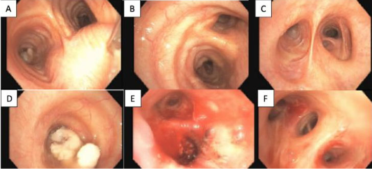
A 74-year-old Caucasian male with a past medical history of alcohol abuse and bilateral inguinal hernias presented with painless jaundice, dark urine, and diarrhea. The patient was a non-smoker. He had a family history of gastric cancer in his mother, heart disease in his father, and prostate cancer in his brother (onset around age 50). Labs revealed elevated alkaline phosphatase of 423 U/L, mildly elevated aspartate aminotransferase and alanine aminotransferase of 53 and 42 U/L, respectively, and an elevated total and direct bilirubin of 18.8 and 15.8 mg/dL, respectively. Abdominal ultrasound revealed intrahepatic and extrahepatic biliary ductal dilation along with diffuse gallbladder wall thickening, gallbladder calculi, and sludge. An irregular pancreatic head mass was discovered on endoscopic ultrasound (EUS), however, fine-needle aspiration biopsies were negative for malignant cells. Biopsy of peri-pancreatic lymph nodes showed atypical cells. Additionally, endoscopic retrograde cholangiopancreatography (ERCP) identified a malignant appearing common bile duct stricture for which he underwent successful sphincterotomy, dilation, and stenting. Carcinoembryonic antigen (CEA) and carbohydrate antigen 19-9 (CA 19-9) levels drawn prior to ERCP were 7.4 ng/mL (reference range 0–5 ng/mL) and 8 U/mL (reference range <34 U/mL), respectively. Computed tomography (CT) imaging of the chest, abdomen, and pelvis revealed mildly enlarged mediastinal, abdominal, and retroperitoneal lymph nodes and patchy sclerosis of several vertebral bodies (Figure 1). Magnetic resonance imaging (MRI) with magnetic resonance cholangiopancreatography (MRCP) showed post-contrast enhancing soft tissue at the hilum of the liver extending into the right and left hepatic ducts concerning for a Klatskin tumor (Figure 2, Figure 3, Figure 4).
Bronchoscopy with endobronchial ultrasound and biopsy of the mediastinal lymph nodes was performed, and pathology of the mediastinal nodes revealed malignant cells with immunohistochemical staining positive for PSA and PSAP and negative for TTF-1, CK7, and CDX2, features consistent with metastatic prostate adenocarcinoma. After this result, PSA was checked and resulted elevated to 205 ng/mL (reference range 0–4 ng/mL). The patient underwent repeat upper EUS and ERCP with biopsy of the bile duct and a gastrohepatic lymph node. The bile duct biopsy was negative for malignant cells; however, the gastrohepatic lymph node pathology was consistent with metastatic prostate adenocarcinoma. The patient’s workup was suspicious for both a Klatskin-type cholangiocarcinoma and metastatic prostate cancer, although biliary brushing samples repeatedly failed to demonstrate findings consistent with primary biliary cancer. The patient had continued workup for the apparent Klatskin tumor, and eventually underwent a liver wedge biopsy, common bile duct excisional biopsy, cholecystectomy, and chest wall port placement in anticipation of chemoradiation for suspected cholangiocarcinoma. Surprisingly, the surgical pathology including from the perihilar lesion was again consistent with metastatic prostate adenocarcinoma.
The patient was referred to medical oncology and began androgen deprivation therapy (ADT) with leuprolide in addition to daily abiraterone with prednisone. Three months after beginning ADT the patient required inpatient admission for biliary sepsis. He improved after undergoing repeat ERCP with biliary stent exchange, and repeat PSA at the time was 2.6 ng/mL. The patient has tolerated leuprolide, abiraterone, and prednisone well overall despite mild fatigue and intermittent hot flashes, and he has experienced marked improvement in quality of life. At the time of manuscript submission, it has been 18 months since the patient first presented with painless jaundice and was diagnosed with metastatic prostate adenocarcinoma. He continues to do well on systemic therapy and presents for repeat PSA and medical oncology follow-up every three months. Serum bilirubin has remained within normal values even after removal of all biliary stents, and his most recent serum PSA value was 0.2 ng/mL.


Discussion
Our patient presented to medical attention due to subacute onset of jaundice, dark urine, and decreased appetite—findings suspicious for biliary obstruction. Initial abdominal ultrasound and CT imaging revealed intrahepatic and extrahepatic biliary ductal dilation without a clear mass in the biliary tract, but subsequent MRI of the pancreas identified a discrete hilar lesion. The findings were most consistent with a Klatskin-type extrahepatic cholangiocarcinoma.
Cholangiocarcinoma is a rare epithelial malignancy that arises from cholangiocytes within the biliary tree. It is anatomically classified as intrahepatic or extrahepatic, and both subtypes have a characteristic clinical presentation, though the presenting symptoms can be overlapping and nonspecific. Intrahepatic cholangiocarcinomas arise in bile ducts located within the liver. These lesions are less likely to lead to jaundice at the time of initial presentation—symptomatic patients more often present with abdominal pain and weight loss [3]. Patients with extrahepatic cholangiocarcinoma more often present with biliary obstruction [4]. Perihilar lesions constitute the majority of extrahepatic cholangiocarcinoma, including those involving the bifurcation of the left and right hepatic ducts, now colloquially known as Klatskin tumors. The now widely used eponym for perihilar cholangiocarcinoma is derived from Dr. Gerald Klatskin’s 1965 publication [5] describing 13 cases of hepatic duct adenocarcinoma with similar clinical, anatomic, and pathologic features. In contrast to cholangiocarcinoma, patients with prostate cancer typically do not develop symptoms of biliary obstruction. Most men newly diagnosed with prostate cancer are asymptomatic and the disease is discovered after PSA screening. Metastatic disease is present upon diagnosis in a minority of patients with prostate cancer, and the most common sites of metastatic disease are the bone, lymph nodes, or liver [2].
There are a number of reports describing patients with imaging findings suspicious for cholangiocarcinoma, later determined attributable to a variety of non-neoplastic entities including tuberculosis, acquired immunodeficiency syndrome (AIDS), and biliary sarcoidosis [6],[7],[8],[9],[10]. Lesions seen in primary sclerosing cholangitis (PSC) can mimic malignancy; a troublesome association considering the lifetime risk of cholangiocarcinoma among patients with PSC is estimated to be between 5% and 13% [11]. There are reports of varied neoplastic conditions imitating Klatskin tumors as well, including malignant melanoma, carcinoid tumors, non-Hodgkin’s lymphoma including diffuse large B-cell lymphoma, and myeloid sarcoma [12],[13],[14],[15],[16].
Descriptions of prostate cancer presenting with clinical and radiographic features of a Klatskin tumor are extremely rare. Published in 1990 in the Annals of Internal Medicine, Chen et al. describe the case of a 77-year-old male who presented with scleral icterus, dark urine, and clay-colored stools [17]. Cross-sectional imaging revealed biliary obstruction at the confluence of the right and left hepatic ducts. Physical exam was notable for an enlarged prostate, but there were no palpable prostatic nodules. Workup revealed elevation in serum prostatic acid phosphatase, a marker utilized routinely in the era prior to PSA screening, and prostate biopsy was performed which yielded a diagnosis of prostate adenocarcinoma. This was believed to be localized initially; however, shortly thereafter an exploratory laparotomy was performed and examination of tissue samples from the hepatic hilum, peritoneum, and gallbladder were all consistent with metastatic well-differentiated adenocarcinoma of the prostate gland. The authors prodigiously surmised “no physician is infallible in the differential diagnosis of obstructive jaundice.” The patient was managed with bilateral orchiectomy, and was asymptomatic 11 months from the time of initial presentation. The finding of an enlarged prostate based on a thorough physical exam led to prostate biopsy early in the workup, whereas in our case, prostate biopsy was deferred entirely. In the setting of confirmed metastatic disease, the potential benefits were not felt to outweigh potential risks of the procedure, and prostate biopsy results were not expected to alter management. We have identified three additional cases in the literature describing metastatic prostate cancer as an etiology for biliary obstruction, though with imaging findings less consistent with Klatskin tumors [18],[19],[20]. Nigam et al. describe the case of a 48-year-old male with an established diagnosis of metastatic prostate cancer with known bone, lung, and widespread lymph node involvement who was found to have biliary obstruction related to metastatic prostate cancer as well, proven with ERCP and biliary sampling [18]. Our case is most similar to that described by Chen et al. [17] and to our knowledge, it is only the second reported case of metastatic prostate cancer presenting with jaundice combined with imaging consistent with Klatskin cholangiocarcinoma.
Our case underscores the importance of definitive pathological tissue diagnosis, a mandatory but often challenging endeavor in the case of biliary cancers. The patient we describe underwent multiple ERCP procedures that were non-diagnostic for malignancy. In addition, the patient had bronchoscopy with mediastinal lymph node sampling, EUS with gastrohepatic lymph node sampling, and finally laparoscopic liver wedge biopsy and excisional biopsy of the affected segment of the common bile duct (CBD), as it was unclear if the patient had a Klatskin tumor in addition to metastatic prostate cancer. In such challenging cases, it is important to objectively evaluate several data points over time, and to avoid anchoring bias, which is the tendency to place excess importance on the initial information available early in the diagnostic workup. Furthermore, this case exemplifies the importance of an interdisciplinary approach to cancer care. Ultimately, an exhaustive diagnostic workup including multiple biopsies was necessary to prove the single unifying diagnosis of metastatic prostate cancer.
He was subsequently started on androgen deprivation therapy (ADT) and abiraterone with prednisone. Frontline ADT has been the mainstay of management of advanced prostate cancer, and several trials have demonstrated positive outcomes by augmenting ADT with chemotherapy or targeted antiandrogen therapies. Based on the improved overall survival data from the CHAARTED trial for the subset of patients with visceral metastases, treating with ADT combined with docetaxel would have been a reasonable first-line approach in our patient. However, in light of the patient’s age, performance status, and status-post major abdominal surgery, chemotherapy was not utilized. We opted to treat with ADT plus abiraterone with prednisone due to proven benefit and favorable side effect profile of this regimen demonstrated by two landmark trials [21],[22].
Conclusion
Prostate adenocarcinoma and cholangiocarcinoma are distinctly different diseases, and patients with metastatic prostate adenocarcinoma have far superior survival rates. Our case demonstrates an unusual presentation of metastatic prostate cancer masquerading as a Klatskin tumor and we highlight how the accurate diagnosis in our patient has led to an excellent clinical response to prostate cancer treatment.
REFERENCES
1.
SEER Cancer Stat Facts: Prostate Cancer. National Cancer Institute. 2022. [Avaliable at: https://seer.cancer.gov/statfacts/html/prost.html]

2.
Gandaglia G, Abdollah F, Schiffmann J, et al. Distribution of metastatic sites in patients with prostate cancer: A population-based analysis. Prostate 2014;74(2):210–6. [CrossRef]
[Pubmed]
3.
Brown KM, Parmar AD, Geller DA. Intrahepatic cholangiocarcinoma. Surg Oncol Clin N Am 2014;23(2):231–46. [CrossRef]
[Pubmed]
4.
Blechacz B, Komuta M, Roskams T, Gores GJ. Clinical diagnosis and staging of cholangiocarcinoma. Nat Rev Gastroenterol Hepatol 2011;8(9):512–22. [CrossRef]
[Pubmed]
5.
Klatskin G. Adenocarcinoma of the hepatic duct at its bifurcation within the porta hepatis. An unusual tumor with distinctive clinical and pathological features. Am J Med 1965;38:241–56. [CrossRef]
[Pubmed]
6.
Menias CO, Surabhi VR, Prasad SR, Wang HL, Narra VR, Chintapalli KN. Mimics of cholangiocarcinoma: Spectrum of disease. Radiographics 2008;28(4):1115–29. [CrossRef]
[Pubmed]
7.
Arora R, Sharma A, Bhowate P, Bansal VK, Guleria S, Dinda AK. Hepatic tuberculosis mimicking Klatskin tumor: A diagnostic dilemma. Indian J Pathol Microbiol 2008;51(3):382–5. [CrossRef]
[Pubmed]
8.
Dolmatch BL, Laing FC, Ferderle MP, Jeffrey RB, Cello J. AIDS-related cholangitis: Radiographic findings in nine patients. Radiology 1987;163(2):313–6. [CrossRef]
[Pubmed]
9.
Pungpapong S, Steers JL, Wallace MB, Krishna M, Keaveny AP. Hepatobiliary sarcoidosis mimicking Klatskin’s cholangiocarcinoma. Gastrointest Endosc 2006;64(1):124–5. [CrossRef]
[Pubmed]
10.
Alam I, Levenson SD, Ferrell LD, Bass NM. Diffuse intrahepatic biliary strictures in sarcoidosis resembling sclerosing cholangitis. Case report and review of the literature. Dig Dis Sci 1997;42(6):1295–301. [CrossRef]
[Pubmed]
11.
Burak K, Angulo P, Pasha TM, Egan K, Petz J, Lindor KD. Incidence and risk factors for cholangiocarcinoma in primary sclerosing cholangitis. Am J Gastroenterol 2004;99(3):523–6. [CrossRef]
[Pubmed]
12.
Wagner MS, Shoup M, Pickleman J, Yong S. Primary malignant melanoma of the common bile duct: A case report and review of the literature. Arch Pathol Lab Med 2000;124(3):419–22. [CrossRef]
[Pubmed]
13.
Maitra A, Krueger JE, Tascilar M, et al. Carcinoid tumors of the extrahepatic bile ducts: A study of seven cases. Am J Surg Pathol 2000;24(11):1501–10. [CrossRef]
[Pubmed]
14.
Eliason SC, Grosso LE. Primary biliary malignant lymphoma clinically mimicking cholangiocarcinoma: A case report and review of the literature. Ann Diagn Pathol 2001;5(1):25–33. [CrossRef]
[Pubmed]
15.
Nguyen GK. Primary extranodal non-Hodgkin’s lymphoma of the extrahepatic bile ducts. Report of a case. Cancer 1982;50(10):2218–22. [CrossRef]
[Pubmed]
16.
González-Vela MC, Val-Bernal JF, Mayorga M, Cagigal ML, Fernández F, Mazorra F. Myeloid sarcoma of the extrahepatic bile ducts presenting as obstructive jaundice. APMIS 2006;114(9):666–8. [CrossRef]
[Pubmed]
17.
Chen MC, Mullane MR. Metastatic prostatic carcinoma presenting as cholangiocarcinoma (“Klatskin tumor”). Ann Intern Med 1990;112(11):881. [CrossRef]
[Pubmed]
18.
Nigam N, Chalhoub W, Abdelaziz D, Sultan M, Marino G. Obstructive jaundice from metastatic prostate cancer: 1038. American College of Gastroenterology 2014;109:S309.
19.
Sarwar R, Youssef W, Uzoaru I. A rare case of metastatic prostate carcinoma presenting as obstructive jaundice: 2106. American College of Gastroenterology 2017;112:S1168–9.
20.
Bloch WE, Block NL. Metastatic prostate cancer presenting as obstructive jaundice. Urology 1992;40(5):456–7. [CrossRef]
[Pubmed]
21.
Fizazi K, Tran N, Fein L, et al. Abiraterone plus prednisone in metastatic, castration-sensitive prostate cancer. N Engl J Med 2017;377(4):352–60. [CrossRef]
[Pubmed]
22.
James ND, de Bono JS, Spears MR, et al. Abiraterone for prostate cancer not previously treated with hormone therapy. N Engl J Med 2017;377(4):338–51. [CrossRef]
[Pubmed]
SUPPORTING INFORMATION
Author Contributions
Michael A Brister - Conception of the work, Design of the work, Drafting the work, Revising the work critically for important intellectual content, Final approval of the version to be published, Agree to be accountable for all aspects of the work in ensuring that questions related to the accuracy or integrity of any part of the work are appropriately investigated and resolved.
Eshaan N Parikh - Conception of the work, Design of the work, Drafting the work, Revising the work critically for important intellectual content, Final approval of the version to be published, Agree to be accountable for all aspects of the work in ensuring that questions related to the accuracy or integrity of any part of the work are appropriately investigated and resolved.
Mark T DiMarcangelo - Conception of the work, Design of the work, Drafting the work, Revising the work critically for important intellectual content, Final approval of the version to be published, Agree to be accountable for all aspects of the work in ensuring that questions related to the accuracy or integrity of any part of the work are appropriately investigated and resolved.
Robert A Somer - Conception of the work, Design of the work, Revising the work critically for important intellectual content, Final approval of the version to be published, Agree to be accountable for all aspects of the work in ensuring that questions related to the accuracy or integrity of any part of the work are appropriately investigated and resolved.
Julia C Shih - Conception of the work, Design of the work, Drafting the work, Revising the work critically for important intellectual content, Final approval of the version to be published, Agree to be accountable for all aspects of the work in ensuring that questions related to the accuracy or integrity of any part of the work are appropriately investigated and resolved.
Guaranter of SubmissionThe corresponding author is the guarantor of submission.
Source of SupportNone
Consent StatementWritten informed consent was obtained from the patient for publication of this article.
Data AvailabilityAll relevant data are within the paper and its Supporting Information files.
Conflict of InterestAuthors declare no conflict of interest.
Copyright© 2022 Michael A Brister et al. This article is distributed under the terms of Creative Commons Attribution License which permits unrestricted use, distribution and reproduction in any medium provided the original author(s) and original publisher are properly credited. Please see the copyright policy on the journal website for more information.


