 |
Case Report
A case report on malignant perivascular epithelioid cell neoplasm of the thigh, a rare presentation in the skin
1 Department of Internal Medicine, The Brooklyn Hospital Center, 121 De Kalb Avenue, Brooklyn, NY 11201, USA
2 Division of Hematology-Oncology, The Brooklyn Hospital Center, 121 De Kalb Avenue, Brooklyn, NY 11201, USA
3 Department of Pathology, The Brooklyn Hospital Center, 121 De Kalb Avenue, Brooklyn, NY 11201, USA
Address correspondence to:
Samridhi Sinha
Department of Internal Medicine, The Brooklyn Hospital Center, 121 De Kalb Avenue, Brooklyn, NY 11201,
USA
Message to Corresponding Author
Article ID: 100083Z10SS2021
Access full text article on other devices

Access PDF of article on other devices


How to cite this article
Sinha S, Rathnakumar GR, Guevara E, Baqui A. A case report on malignant perivascular epithelioid cell neoplasm of the thigh, a rare presentation in the skin. J Case Rep Images Oncology 2021;7:100083Z10SS2021.ABSTRACT
Perivascular epithelioid cell neoplasms (PEComa) are a family of mesenchymal tumors characterized by perivascular epithelioid cells with co-expression of melanocytic and muscle markers and can occur in any part of the body. These tumors are almost always positive for HMB-45 and smooth muscle actin (SMA) immunochemistry. Perivascular epithelioid cell neoplasms tend to arise in middle age, and there is a marked female predominance. The tumors most commonly occur in the gynecologic tract, pelvis, and retroperitoneum. They are of varying malignant potential and are occasionally associated with tuberous sclerosis complex, a rare tumor driven by tuberous sclerosis complex gene mutations TSC1/2, causing up-regulation of mTOR. We present a case of malignant perivascular epithelioid cell neoplasm of the thigh, which is an exceedingly rare presentation in the skin.
Keywords: Epithelioid features, HMB-45, Melan-A, Mesenchymal tumors, PEComa, Perivascular, Perivascular epithelioid cell neoplasms, Wide resection
Introduction
Perivascular epithelioid cell neoplasms (PEComa) are a family of related mesenchymal tumors characterized by the presence of perivascular epithelioid cells with co-expression of melanocytic (HMB-45 and/or melan A) and smooth muscle markers (actin and/or desmin). Perivascular epithelioid cell neoplasms (PEComa) are histologically and immunophenotypically similar to mesenchymal neoplasms including clear cell sugar tumor (CCST) of the lungs, angiomyolipoma, lymphangioleiomyomatosis.
Case Report
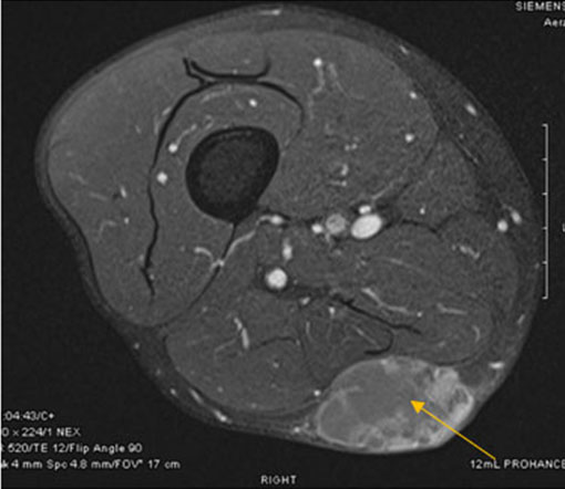
A previously healthy 79-year-old male presented with complaints of one-year history of increasing mass on the posterior right thigh. The patient denied fever, weight loss, and night sweats, or other type B symptoms. Additional review of the system was negative. The patient was diagnosed with 5.5 cm exophytic subcutaneous tumor of the right posterior distal thigh. Complete blood count with automated differentials and comprehensive metabolic profile were within the normal range. Computed tomography (CT) with intravenous contrast showed a low density, partially solid, and cystic mass (Figure 1).
The patient underwent surgical resection with a biopsy of 4.5 × 4.2 cm well-defined nodule with both cystic and solid area, with a solid area measuring 2.5 × 2.5 cm.
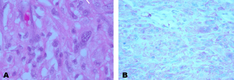
Hematoxylin and eosin staining (Figure 2A and Figure 2B) and immunohistochemistry were performed on the formalin-fixed, paraffin-embedded tissue section using a panel of the following antibodies: vimentin, CD19, SMA, desmin, and cathepsin, HMB45, CK, 10, calretinin, CD 31, CD45, CD68, and CD34.
Our case revealed a superficial, mostly encapsulated high-grade spindle and pleomorphic sarcoma showing large cystic areas. The tumor was composed of the spindle to plump ovoid cells with abundant eosinophilic to vacuolated cytoplasm with no lymph-vascular invasion (Figure 2A and Figure 2B). Upon immunohistochemical staining, the cells were positive for vimentin, CD19, SMA, and desmin, while negative for CK10, calretinin, CD 31, CD45, CD68, CD34, which ruled out epithelioid angiosarcoma. Immunostaining performed in our lab show diffuse and strong desmin and cathepsin K positivity and strong focal reactivity for HMB-45. Simultaneously, other stains such as myogenin, CDK4, MDM2, SOX10, and Melan A are all negative. Stains for TFE3 and SATB2 show some weak/equivocal nuclear staining. Although the positivity for desmin and SMA suggested a diagnosis of leiomyosarcoma, the co-expression of HMB45 and cathepsin K is more in keeping with a diagnosis of malignant perivascular epithelioid cell neoplasms. Fluorescence in situ hybridization (FISH) studies using custom bacterial artificial chromosome (BAC) probes showed no TFE3 gene abnormalities.
Overall findings confirmed malignant perivascular epithelioid cell neoplasms. According to Fédération Nationale des Centres de Lutte Contre Le Cancer (FNCLCC) grading tumor was Grade 3 with the staging of pT1Nx.
The post-operative course was complicated by seroma, which was surgically drained and treated with intravenous antibiotics. No signs of recurrence were noted so far in the interval follow-up. The case was discussed in the tumor board, and sirolimus was decided to be the adjuvant therapy of choice in case of metastatic recurrence.
Discussion
Although perivascular epithelioid cell neoplasms were first described in 1943 by Apitz [1] (as an “abnormal myoblast” in renal angiomyolipoma; it was in 1996 that Zamboni et al. in their first case of pancreatic clear-cell “sugar” tumor (CCST) suggested the name PEComa for neoplasms composed of a pure proliferation of perivascular epithelioid cells [2].
As per the World Health Organization classification of tumors of soft tissues and bones, perivascular epithelioid cell neoplasms are classified as mesenchymal tumors with a make-up of perivascular epithelioid cells [3].
PEComas occasionally associated with tuberous sclerosis complex, are defined by the presence of perivascular epithelioid cells that co-express muscle and melanocytic markers. This family of tumors includes angiomyolipoma (AML), clear cell sugar tumor of the lung (CCST), lymphangioleiomyomatosis (LAM), and very rare tumors in other locations [4].
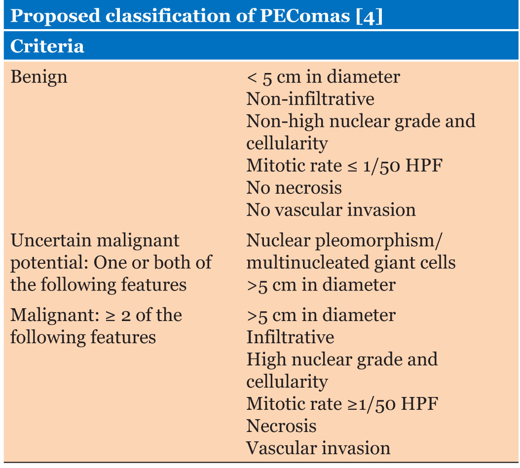
Immunohistochemically angiomyolipomas, lymphangiomyomas, and related tumors are distinctive and co-express markers of smooth muscle (consistently SMA-positive, variably desmin-positive) and melanocytes (HMB-45, Melan-A, MART). Therefore, such a constellation of markers is useful in the diagnosis of angiomyolipoma-related mesenchymal tumors, especially when they occur in an unusual clinical setting [5],[6]. Renal AML is the most common PEComa, with a prevalence of 0.13%, PEComas at other sites, such as soft tissue, bone, visceral, and gynecological sites, are very rare, and occur mainly in middle-aged females with male to female ratio of 1:7 [4]. Folpe et al. proposed the categorization of PEComas into three groups based on tumor diameter, nuclear grade and cellularity, mitotic rate, necrosis, vascular invasion, and infiltrative growth [4] (Table 1).
The final diagnosis of PEComa depends on pathology and immunohistochemistry [3],[7]. PEComas are characterized by their perivascular location, and the cells are with radial arrangement around the vascular lumen. Typically, cells around the vessels are epithelioid and spindle-shaped, resembling smooth muscles cell, and have abundant clear to eosinophilic granular cytoplasm [8],[9].
Immunohistochemical stains play a major role in the diagnosis of PEComas. These tumors almost always co-express SMA and HMB45; over 50% of them stain for melan-A (sometimes in the absence of HMB45), and around 25% are desmin positive. A subset of cases stain for S-100 protein (allowing possible confusion with melanoma) and TFE3 [4].
Conclusion
Perivascular epithelioid cell neoplasms of the skin are rare tumors and are usually seen in the gynecologic tract, pelvis, and retroperitoneum. These tumors are almost always positive for HMB-45/Melan A and SMA stains. The mainstay of treatment is surgical resection for primary tumors and in cases of local recurrence. Close surveillance clinically accompanied by radiological imaging is mandatory. mTOR inhibitors like sirolimus may be considered as adjuvant chemotherapy in patients with recurrence.
REFERENCES
1.
Apitz K. Die Geschwülste und Gewebsmissbildungen der Nierenrinde. II. Midteilung. Die mesenchymalen Neubildungen. Virchows Archiv 1943;311:306–27.

2.
Zamboni G, Pea M, Martignoni G, et al. Clear cell “sugar” tumor of the pancreas. A novel member of the family of lesions characterized by the presence of perivascular epithelioid cells. Am J Surg Pathol 1996;20(6):722–30. [CrossRef]
[Pubmed]
3.
Fletcher CD, Unni KK, Mertens F. World Health Organization Classification of Tumors of Pathology and Genetics of Tumours of Soft Tissue and Bone. Lyon: IARC Press; 2002. p. 221–2.
4.
Folpe AL, Mentzel T, Lehr HA, Fisher C, Balzer BL, Weiss SW. Perivascular epithelioid cell neoplasms of soft tissue and gynecologic origin: A clinicopathologic study of 26 cases and review of the literature. Am J Surg Pathol 2005;29(12):1558–75. [CrossRef]
[Pubmed]
5.
Bonetti F, Pea M, Martignoni G, et al. Clear cell (“sugar”) tumor of the lung is a lesion strictly related to angiomyolipoma – the concept of a family of lesions characterized by the presence of the perivascular epithelioid cells (PEC). Pathology 1994;26(3):230–6. [CrossRef]
[Pubmed]
6.
Fetsch PA, Fetsch JF, Marincola FM, Travis W, Batts KP, Abati A. Comparison of melanoma antigen recognized by T cells (MART-1) to HMB-45: Additional evidence to support a common lineage for angiomyolipoma, lymphangiomyomatosis, and clear cell sugar tumor. Mod Pathol 1998;11(8):699–703.
[Pubmed]
7.
Patra S, Vij M, Kota V, Kancherla R, Rela M. Pigmented perivascular epithelioid cell tumor of the liver: Report of a rare case with brief review of literature. J Cancer Res Ther 2013;9(2):305–7. [CrossRef]
[Pubmed]
8.
Akitake R, Kimura H, Sekoguchi S, et al. Perivascular epithelioid cell tumor (PEComa) of the liver diagnosed by contrast-enhanced ultrasonography. Intern Med 2009;48(24):2083–6. [CrossRef]
[Pubmed]
9.
Folpe AL, Kwiatkowski DJ. Perivascular epithelioid cell neoplasms: Pathology and pathogenesis. Hum Pathol 2010;41(1):1–15. [CrossRef]
[Pubmed]
SUPPORTING INFORMATION
Author Contributions
Samridhi Sinha - Conception of the work, Design of the work, Acquisition of data, Analysis of data, Drafting the work, Final approval of the version to be published, Agree to be accountable for all aspects of the work in ensuring that questions related to the accuracy or integrity of any part of the work are appropriately investigated and resolved.
Geethapriya Rajasekharan Rathnakumar - Conception of the work, Design of the work, Acquisition of data, Analysis of data, Revising the work critically for important intellectual content, Final approval of the version to be published, Agree to be accountable for all aspects of the work in ensuring that questions related to the accuracy or integrity of any part of the work are appropriately investigated and resolved.
Elizabeth Guevara - Conception of the work, Design of the work, Acquisition of data, Analysis of data, Drafting the work, Revising the work critically for important intellectual content, Final approval of the version to be published, Agree to be accountable for all aspects of the work in ensuring that questions related to the accuracy or integrity of any part of the work are appropriately investigated and resolved.
Aam Baqui - Conception of the work, Design of the work, Acquisition of data, Analysis of data, Drafting the work, Revising the work critically for important intellectual content, Final approval of the version to be published, Agree to be accountable for all aspects of the work in ensuring that questions related to the accuracy or integrity of any part of the work are appropriately investigated and resolved.
Guaranter of SubmissionThe corresponding author is the guarantor of submission.
Source of SupportNone
Consent StatementWritten informed consent was obtained from the patient for publication of this article.
Data AvailabilityAll relevant data are within the paper and its Supporting Information files.
Conflict of InterestAuthors declare no conflict of interest.
Copyright© 2021 Samridhi Sinha et al. This article is distributed under the terms of Creative Commons Attribution License which permits unrestricted use, distribution and reproduction in any medium provided the original author(s) and original publisher are properly credited. Please see the copyright policy on the journal website for more information.


