 |
|
Case Report
| ||||||
| A rare case of primary pulmonary angiosarcoma | ||||||
| Joel Alcid1, Wayne Pinover2 | ||||||
|
1Resident, Internal Medicine, Drexel, Philadelphia, PA, USA 2Attending, Radiation Oncology, Abington-Jefferson Health, Abington, PA, USA | ||||||
| ||||||
|
[HTML Abstract]
[PDF Full Text]
[Print This Article]
[Similar article in Pumed] [Similar article in Google Scholar] |


| How to cite this article |
| Alcid J, Pinover W. A rare case of primary pulmonary angiosarcoma. J Case Rep Images Oncology 2018;4:100047Z10JA2018. |
|
ABSTRACT
|
|
Introduction: Angiosarcoma is a malignant epithelial cell neoplasm of vascular or lymphatic origin. The key features of behavior of these tumors include insidious growth, extensive local invasion and hematogenous metastasis. Involvement of the lung usually indicates metastatic disease. Very few cases of primary pulmonary angiosarcoma have been reported. We present a case of a 77-year-old female with primary pulmonary angiosarcoma. Case Report: A 77-year-old female, with a 30-pack year smoking history and current half pack per day smoker, presented to the emergency department with progressive shortness of breath and hemoptysis. Patient had seen her primary care provider one week prior to arrival and was advised to have a computed tomography scan of the chest, which showed 8x4.6x10.4 cm cystic/necrotic septated mass in the left posterior mediastinum. She was started on apixaban for this questionable tumor extension versus bland clot. Hemoptysis had become significantly heavier since starting apixaban, so she came to the emergency department for further evaluation. A fine needle aspiration (FNA) with endobronchial ultrasound (EBUS) was performed, histological examination of the biopsy specimen showed a high grade spindled and epithelioid malignant cells with associated blood vessels and extensive necrosis. Immunohistochemical staining of the tumor revealed it was strongly positive for CD31, vimentin, and cytokeratin, suggestive of epithelioid angiosarcoma. After five doses of radiation, patient was discharged to short term rehab facility. Based on the progression and aggressiveness of the tumor, the patient decided to go on hospice. Shortly after, the patient died from respiratory failure. Conclusion: Primary pulmonary angiosarcoma is a rare malignant vascular tumor which usually represents metastasis from another primary site. Only a few cases of primary pulmonary angiosarcoma have been reported in literature. Prognosis is poor, with mortality within the first few months of presentation approaching 100%. | |
|
Keywords: Oncology, Primary, Pulmonary angiosarcoma, Radiation | |
|
INTRODUCTION
| ||||||
|
Primary pulmonary angiosarcoma is a rare type of malignant vascular tumor. Angiosarcoma of the lung usually represents metastasis from another primary site. The primary tumors are most commonly found in the head/neck region, and may frequently metastasize to the lung. The key features of behavior of these tumors include insidious growth, extensive local invasion and hematogenous metastasis. No standard treatment regimen has been established for pulmonary angiosarcoma. Radiation therapy, chemotherapy, surgical resection, and immunotherapy have been attempted. A primary lung tumor is extremely rare, with only a several cases reported. | ||||||
|
CASE REPORT
| ||||||
|
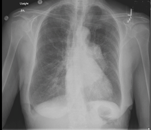
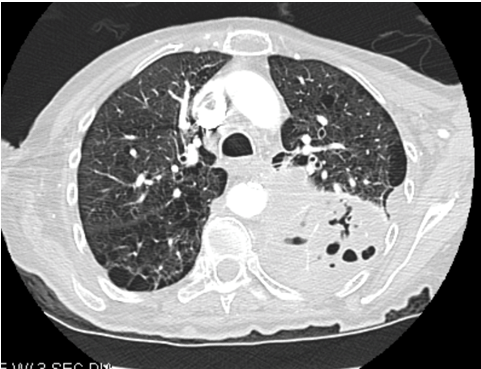
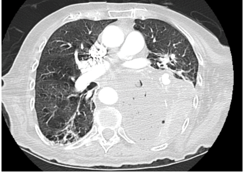
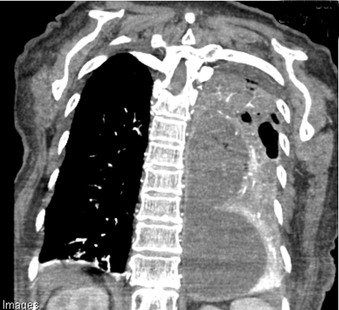
A 77-year-old female, with a 30-pack year smoking history and current 1/2 pack per day smoker, presented to the emergency department with progressive shortness of breath and hemoptysis. The patient had seen her primary care provider one week prior to arrival and was advised to have a computed tomography scan of the chest, which showed 8x4.6x10.4 cm cystic/necrotic septated mass in the left posterior mediastinum. There was also mass effect of adjacent structures including the left lower pulmonary vein which demonstrated a thrombus within the lumen (Figure 1) (Figure 2). She was started on apixaban for this questionable tumor extension versus bland clot. Hemoptysis had become significantly heavier since starting Eliquis, so she came to the emergency department for further evaluation. A fine needle aspiration (FNA) with endobronchial ultrasound (EBUS) was performed, and after removal of a mucus plug in the left lower lobe, brisk bleeding occurred. Hemostasis was achieved with an endobronchial blocker and biopsy of the mass was taken. Histological examination of the biopsy specimen showed a high grade spindled and epithelioid malignant cells with associated blood vessels and extensive necrosis. Immunohistochemical staining of the tumor revealed it was strongly positive for CD31, vimentin, and cytokeratin, suggestive of epithelioid angiosarcoma. Imaging did not reveal additional sites of malignancy, and a diagnosis of primary pulmonary angiosarcoma was made. The patient was not a surgical candidate based on the size and complexity of the tumor, along with the poor performance status. Radiation oncology was consulted for further management and radiation treatments were started. After five-doses of radiation, patient was discharged to short-term rehab facility. Within one month of discharge, patient returned to the emergency department with worsening cough, shortness of breath, hemoptysis and performance status. The patient received 65% of planned radiation treatments prior to arrival. Computed tomography scan of the chest revealed a 14x9x6 cm large mass like structure in the left lower lobe containing complex fluid and multiple pockets of air suggestive of an infected or necrotic tumor (Figure 3) (Figure 4) (Figure 5). Based on the progression and aggressiveness of the tumor, the patient decided to go on hospice. Shortly after, the patient died from respiratory failure. | ||||||
| ||||||
| ||||||
| ||||||
|
| ||||||
|
| ||||||
|
DISCUSSION
| ||||||
|
Primary pulmonary angiosarcoma is a rare type of malignant vascular tumor. Angiosarcoma of the lung usually represents metastasis from another primary site. The primary tumors are most commonly found in the head/neck region, and may frequently metastasize to the lung. However, a primary lung tumor is extremely rare, with only a several cases reported [1]. Patients present with nonspecific respiratory symptoms which include hemoptysis, dyspnea, cough, chest pain, and weight loss. Early diagnosis is uncommon, because of its rarity and low index of suspicion. The average age of diagnosis according to the data in literature is 55.9 years and males are more often affected than females [2][3]. The key features of behavior of these tumors include insidious growth, extensive local invasion and hematogenous metastasis. Definitive diagnosis is made by biopsy and immunohistochemical analysis. Pulmonary angiosarcoma is characterized by the proliferation of tumor cells with endothelial features. Features include variably sized vascular channels with irregular branching, epithelioid like endothelial lining of vascular spaces, abundant cytoplasm with pleomorphic nuclei and prominent nucleoli. Endothelial markers are typically expressed in angiosarcoma. These markers include CD 31, CD 34, and factor VIII related antigen. Of these markers, CD 31 is relatively specific and extremely sensitive, being detected in approximately 90% of cases. In approximately 30% of cases, angiosarcoma expresses cytokeratin, as in our patient [1][4]. No standard treatment regimen has been established for pulmonary angiosarcoma. Radiation therapy, chemotherapy, surgical resection, and immunotherapy have been attempted. For patients with localized disease, surgery has been shown to be the most effective. It is important that surgery be considered as early as possible if the tumor is deemed resectable. Two chemotherapy regimens that have demonstrated full and partial effects are doxorubicin/ifosfamide and docetaxel/gemcitabine. Immunotherapy with recombinant interleukin 2 in combination with either surgery or chemotherapy has also shown to be effective. The use of radiation therapy in conjunction with surgery, has resulted in local control and excellent functional and cosmetic outcome, for patients with angiosarcoma of the head and neck, but not the lung [3] [4][5][6] [7] [8]. | ||||||
|
CONCLUSION
| ||||||
|
Primary pulmonary angiosarcoma is a rare malignant vascular tumor which usually represents metastasis from another primary site. Only a few cases of primary pulmonary angiosarcoma have been reported in literature. Prognosis is poor, with mortality within the first few months of presentation approaching 100%. A high index of suspicion is essential to its early diagnosis and could improve its poor prognosis. | ||||||
|
REFERENCES
| ||||||
| ||||||


|
[HTML Abstract]
[PDF Full Text]
|
|
Author Contributions
Joel Alcid – Substantial contributions to conception and design, Acquisition of data, Drafting the article, Revising it critically for important intellectual content, Final approval of version to be published Wayne Pinover – Substantial contributions to conception and design, Acquisition of data, Drafting the article, Revising it critically for important intellectual content, Final approval of version to be published |
|
Guarantor of Submission
The corresponding author is the guarantor of submission. |
|
Source of Support
None |
|
Conflict of Interest
Authors declare no conflict of interest. |
|
Copyright
© 2018 Joel Alcid et al. This article is distributed under the terms of Creative Commons Attribution License which permits unrestricted use, distribution and reproduction in any medium provided the original author(s) and original publisher are properly credited. Please see the copyright policy on the journal website for more information. |