| Table of Contents |  |
|
Case Report
| ||||||
| Primary mediastinal choriocarcinoma in a male patient: A case report | ||||||
| Ayman A. Rasmy1, Waleed M. Selwi1, Samir T. Fotih1, Ehab A. Hassan1, Usama H. Ismail2 | ||||||
|
1Assistant Consultant, Adult Oncology Department, King Fahad Specialist Hospital, Dammam, KSA.
2Assistant Consultant Cardiology, King Fahad Specialist Hospital, Dammam, KSA. | ||||||
| ||||||
|
[HTML Abstract]
[PDF Full Text]
[Print This Article]
[Similar article in Pumed] [Similar article in Google Scholar] |
| How to cite this article: |
| Rasmy AA, Selwi WM, Fotih ST, Hassan EA, Ismail UH. Primary mediastinal choriocarcinoma in a male patient: A case report. J Case Rep Images Oncology 2015;1:1–4. |
|
Abstract
|
|
Introduction:
Choriocarcinoma is a malignant germ cell neoplasm. It can exhibit in a gonadal site or, rarely, in extra-gonadal sites. Primary mediastinal choriocarcinoma can occur in both male and female in second and third decades of their life, but it is more common in male. The most common symptoms are cough, chest pain and gynecomastia. Plasma chorionic gonadotrophin (β-hCG), which is used as a tumor marker for diagnosis, staging and monitoring of the treatment response. Chemotherapy can achieve response if the case diagnosed early and receive the proper treatment earlier. BEP regimen is the standard first line chemotherapy, unless there is no contraindication specially for the presence of lung disease (Bleomycin) or kidney disease (cisplatin).
Case Report: We describe here a case of a 22-year-old heavy smoker Saudi male for more than 10 years, who was referred to us as a case of advanced pulmonary choriocarcinoma which confirmed by pathology and tumor marker. Our patient was admitted to ICU initially due to shortness of breath. The diagnosis was confirmed as a case of primary mediastinal choriocarcinoma. It could not be treated by surgery due to bad general condition of the patient with severe difficulty in breating requiring respiratory support and ICU care. The chemotherapy was started by VeIP Protocol for one cycle but due to no subjective response as well as the presence of same high level β-hCG, the chemotherapy was changed to TIP Protocol. On the last day of chemotherapy, the patient started to complain about increasing tachypnea and tachycardia. He was transfered to ICU and was was intubated there. Although there was a significant decrease in his β-hCG level, but unfortunately the patient died with multi-organ failure. Conclusion: Early diagnosis of germ cell tumor can increase the chance of cure. The presence of primary disease in different extra-gonadal sites like primary medicinal choriocarcinoma that leads to delay in the diagnosis, which reflected on the treatment outcome with poor overall prognosis even with the presence of different chemotherapy protocols. | |
|
Keywords:
Choriocarcinoma, Extra gonadal Choriocarcinoma, Primary Mediastinal Choriocarcinoma, TIP protocol, VeIP protocol
| |
|
Introduction
| ||||||
|
Testicular cancer constitutes about 1% of cancers in males, germ cell tumors (GCTs) account about 93% of all primary testicular malignancies. Choriocarcinoma is a malignant germ cell neoplasm that accounts less than 1% of all testicular tumors, it is a common component of other testis tumors. Choriocarcinnoma can exhibit in a gonadal site or, rarely, in extra-gonadal sites. Choriocarcinomas are the most aggressive and rapidly growing germ cell tumors. They spread via blood and lymphatic, with early blood dissemination to the lungs, liver and brain. When there is a detectable lesion in the mediastinum, without the presence of the primary lesion in the gonads or metastatic disease in the retroperitoneal lymph nodes, is termed a primary choriocarcinoma of the mediastinum. The primary mediastinal choriocarcinoma in a male is an extremely rare variant commonly between the age 15 to 35 years, with an incidence of 2.1 cases per 100,000 males with four folds higher incidence in white males than in black males. Its syncytiotrophoblastic cells contain plasma chorionic gonadotrophin (β-hCG), which is used as a tumor marker for diagnosis, staging and monitoring of the treatment response. | ||||||
|
Case Report
| ||||||
|
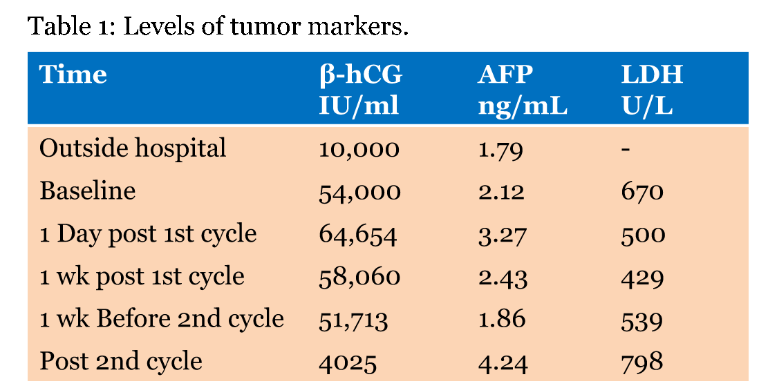
A 22-year-old male patient was referred to our hospital from outside hospital, as a case of germ cell tumor of lung origin according to report from there. He was a heavy smoker for last 10 years, presented there with cough most of the time, which was dry for 3 months, with dyspnea on lying down. The symptoms progressively increased with coughing small quantity of fresh blood with sputum associated with right chest pain with no fever. He was dyspnic, oxygen saturation was 92 to 94% on 10 L of oxygen, heart rate was 102 beats per minute with no lymphadenopathy, no lower limb edema. Chest with stony dullness to percussion noticed with decreased air entry on right side with no organmegaly. Testicular examination was normal with no significant mass. The initial laboratory assessment at that time showed normal complete blood count, and biochemistry. The tumor marker showed high beta-hCG, (more than 10,000 IU/ml) with normal alpha-fetoprotein. The CT chest showed large right side mediastinal mass, about 10x8x15 cm with adjacent compression of superior vena cava and right pulmonary artery with no evidence of invasion and also, there were multiple pulmonary nodules with small right side pleural effusion (Figure 1). Bronchoscopy was done, but the biopsy and BAL was negative for malignant cells. The BAL culture revealed the growth of Escherichia coli and started on antibiotics, according to sensitivity for 6 days, but with no clinically and radiologically improvement, so fine-needle aspiration (FNA) and a biopsy was done from mass and initial report showed it is a germ cell tumor, so the patient was referred to our hospital for further assessment. In our hospital, initially, this patient was admitted in the ICU because of severes hortness of breath, the review of CT done outside showed same finding that was mentioned in the report from outside hospital. Once the diagnosis was confirmed as choriocarcinoma (tumor cells were reactive to HCG and AE1/3 but negative for PLAP, CD30, CD117, AFP and Oct-4), the chemotherapy was started in form of VeIP protocol every 3 weeks as follows: Vinblastine(0.11 mg/kg IV in 50 ml NS over 15 minutes- day-1 and 2), Ifosfamide – (1500 mg/m2 IV in 500 ml D5 1/2 NS over 1 hour. Day 1–4 – with mesna), and Cisplatin (20 mg/m2 IV in 100 ml NS over 30 min – day 1–5). Our decision at hat time based on that we cannot start on BEP protocol, which contain Bleomycin/Etopside/cisplatin because of high risk of pulmonary toxicity with bleomycin, as this patient had an extensive lung disease and heavy smoker. After finishing cycle 1 of chemotherapy, he felt better with less oxygen dependent than before. Portable chest X-ray was done at that time and showed some improvement (Figure 2), so the patient discharged with G-CSF SQ injection daily for 5 days.The baseline β-hCG was 54.000 IU/ml with elevated LDH (574 units/L) and normal fetoprotein (2.12 ng/ml) (Table 1). The patient was staying at home suitable for about 1 week, after that he came to the emergency room with chest pain, his ECG showed a picture of pericarditis and by echocardiogram, there was a compression of mediastinal mass compression the right atrium, so he was admitted to the CCU for 48 hours, started on colchicine and symptomatic treatment with clinical improvement and CT chest with contrast was done at that time and showed some improvement (Figure 3), so he discharged stable. After discharge by one week, the patient admitted again from the emergency with shortness of breath, spiral CT scan of chest was done to rule out pulmonary embolism that was negative, but there was evidence of disease progression in comparison to the previous study (Figure 4). At the time of 2nd cycle, The tumor marker was in same high range as before (β-hCG was 58.083 IU/ml), so bed side tumor board discussion was carred out and final decision was to switch chemotherapy from VeIP to TIP protocol (paclitaxel 175 mg/m2 over 3 hours day 1 with dexamethasone preperation, ifosfamide 1200 mg/m2 over 1 hour day 2–6 with mesna and cisplatin 20 mg/m2 over 30 min with good hydration day 2–6) and the patient started to receive chemotherapy but on 5th day of chemotherapy, he started to complaintfrom tachypnea and tachycardia, , so he was transferred to ICU in which he was intubated and left intercostal tube was inserted there because of the presence of pneumothorax , the filgrastim subcutenous daily injection was started , but unfortunately there was clinical detoriratation and the patient was died on 2nd week in the ICU with multi-organ failure. | ||||||
| ||||||
| ||||||
| ||||||
| ||||||
| ||||||
|
Discussion
| ||||||
|
Germ cell tumors, despite their name, it can occur either inside or outside testis and if it occurred outside testis , it named extragonadal germ cell tumor. Patients often present initially with acute disorders resulting from hemorrhage or necrosis of the primary tumours or their metastases because the average diagnostic delay is 4 to 6 months after symptom onset, published case studies show that choriocarcinoma may present with 1 or more of the following complications. The β-hCG is the standard tumor marker used for diagnosis and for the monitoring of response as well as it can used for follow-up. There is are many theories explaining the origin of choriocarcinoma because the exact cause is unknown. The primitive germ cell theory has received wider acceptance.These cells may arise in the covering mesothelium of the primitive gonad or may arise from the yolk sac endoderm, normally migrating along the urogenital ridge coming to rest in the gonad. In cases of primary mediastinal choriocarcinoma, the arrest of the germ cell somewhere along the path may have occurred. These cells may remain dormant until puberty or later sex life when some stimulus might cause them to mature and develop into a tumor mass [1]. A noteworthy feature of mediastinal choriocarcinoma is that it has a distinctly poorer prognosis than its testicular counterpart [2]. Chemotherapy had a good results when used earlier in the treatment of choriocarcinoma. BEP regimen is the standard first line chemotherapy, unless there is no contraindication specially for the presence of lung disease (Bleomycin) or kidney disease (cisplatin). Other salvage chemotherapy like TIP or VeIP have less response rates. According to Kathuria et al. , this combination has shown some promise in improving survival up to two years after surgery and chemotherapy in mediastinal choriocarcinoma [3]. | ||||||
|
Conclusion
| ||||||
|
Primary medistinal choricarcinoma is one of germ cell tumors that occurred in young age male and need early diagnosis and proper management, otherwise it will be fatal. | ||||||
|
Acknowledgements
| ||||||
|
To all of Oncology team and ICU team for their effort in management of this critical patient. | ||||||
|
SUGGESTED READING
| ||||||
| ||||||
|
References
| ||||||
| ||||||
|
[HTML Abstract]
[PDF Full Text]
|
|
Author Contributions
Ayman A. Rasmy – Substantial contributions to conception and design, Acquisition of data, Analysis and interpretation of data, Drafting the article, Critical revision of the article, Final approval of the version to be published Waleed M. Selwi – Analysis and interpretation of data, Drafting the article, Final approval of the version to be published Samir T. Fotih – Analysis and interpretation of data, Drafting the article, Final approval of the version to be published Ehab A. Hassan – Analysis and interpretation of data, Drafting the article, Final approval of the version to be published Usama H. Ismail – Analysis and interpretation of data, Drafting the article, Final approval of the version to be published |
|
Guarantor of submission
The corresponding author is the guarantor of submission. |
|
Source of support
None |
|
Conflict of interest
Authors declare no conflict of interest. |
|
Copyright
© 2015 Ayman A. Rasmy et al. This article is distributed under the terms of Creative Commons Attribution License which permits unrestricted use, distribution and reproduction in any medium provided the original author(s) and original publisher are properly credited. Please see the copyright policy on the journal website for more information. |
|
|


