| Table of Contents |  |
|
Case Report
| ||||||
| Glassy cell carcinoma arising in the vagina: A case in a young patient | ||||||
| Maher Slimane1, Nada Belhaj Yahia2, Hanene Bouaziz2, Salma Gadria2, Monia Hechiche3, Khaled Rahal4 | ||||||
|
1Assistant, Oncologic Surgery Department, Institute Salah Azaiez, 1006 Tunis, Tunisia.
2Resident, Oncologic Surgery Department, Institute Salah Azaiez, 1006 Tunis, Tunisia. 3Professor, Oncologic Surgery department, Institute Salah Azaiez, 1006 Tunis, Tunisia. 4Department head, Oncologic Surgery Department, Institute Salah Azaiez, 1006 Tunis, Tunisia. | ||||||
| ||||||
|
[HTML Abstract]
[PDF Full Text]
[Print This Article]
[Similar article in Pumed] [Similar article in Google Scholar] |


| How to cite this article: |
| Slimene M, Belhaj Yahia N, Bouaziz H, Gadria S, Hechiche M, Rahal K. Glassy cell carcinoma arising in the vagina: A case in a young patient. J Case Rep Images Oncology 2016;2:28–31. |
|
Abstract
|
|
Introduction:
Glassy cell carcinoma is rare entity especially reported in cervix. Its occurrence in the vagina is exceptional. This article is the second known case of glassy cell carcinoma arising in the vagina. Extrapolating the results related to glassy cell carcinoma of the cervix and data on tumors of the vagina, has allowed us to better define the treatment of this tumor and predict its prognosis. In fact, glassy cells carcinoma is a histologically subtype of cancer known for its loco-regional aggressiveness and the high frequency of metastases. So, the treatment strategy should be discussed in a multidisciplinary meeting.
Case Report: A 24-year-old female patient presented with atypical genital bleeding for about six months and whose different explorations have concluded that it was glassy cell carcinoma arising in the vagina with clinical staging of 'International Federation of Obstetrics and Gynecology' III disease. This case illustrates the potential for glassy cell carcinoma to arise throughout the female genital tract but mainly through the lymphatic node. The patient was successfully treated by neoadjuvant chemotherapy and concomitant radio-chemotherapy proceeded by ovarian transposition. Despite the poor prognosis of this rare entity, after a decline of six months, there was a good therapeutic response with complete disappearance of the tumor. Conclusion: The aggressiveness of the glassy cell carcinoma, rare tumor which can affect the vagina, makes the multidisciplinary management essential. Despite the combination of radiotherapy, chemotherapy and surgery in the treatment of these tumors, their prognosis remains poor. | |
|
Keywords:
Adenosquamous carcinoma, Genital bleeding, Glassy cell carcinoma, Vaginal cancer
| |
|
Introduction
| ||||||
|
Glassy cell carcinoma is considered as the most poorly differentiated form of adenosquamous carcinoma. The most frequent location is in the cervix [1][2][3]. It occurs in various other sites of genital tract: Fallopian tubes, body of endometria and vagina [4][5][6][7]. The management of this tumor is based on surgery, radiotherapy, and chemotherapy [2][3]. To our knowledge, this case is the second reported case of glassy cell carcinoma of the vagina. | ||||||
|
Case Report
| ||||||
|
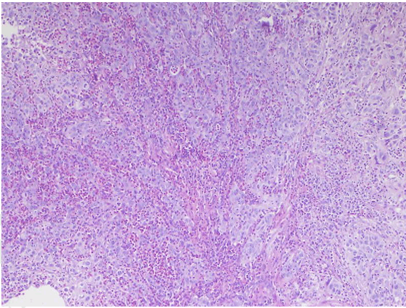
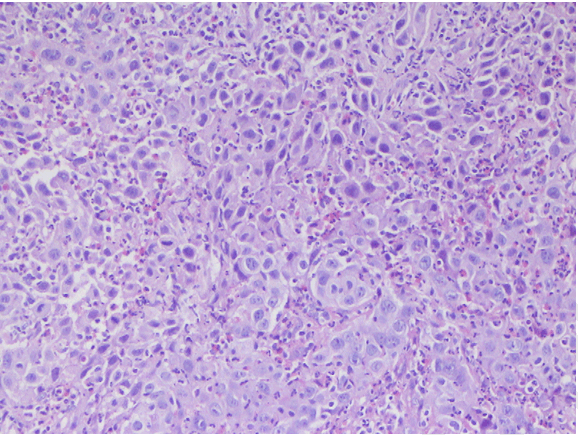
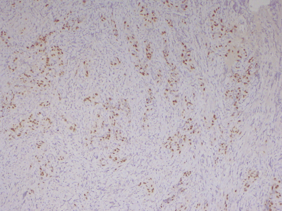
A 24-year-old female was admitted to our department for atypical genital bleeding for about six months. She had no history of prenatal diethylstilbestrol exposure or sexual intercourse. Gynecological examinations revealed a circumscribed, ulcerated tumor originating in the vaginal wall. This tumor was occupied the upper and middle third of vaginal wall. The uterine cervix and parameters were apparently intact. Inguinal lymph nodes were not evident. Microscopic examination of the biopsied specimen of the tumor revealed sheets of large cells with abundant inflammatory infiltration consisting predominantly of eosinophils and lymphocytes characterized by large nuclei with prominent nucleoli. Cells showed a moderate amount of granular or clear cytoplasm. We observed a high mitotic rate and many abnormal mitoses and distinct cell membrane stained well by PAS stain. Finally, the presence of necrosis area was noted. The pathological diagnosis of glassy cell carcinoma was made (Figure 1) (Figure 2) (Figure 3). Abdominal and pelvic computed tomography (CT) scan showed an important tissue thickening affecting all vaginal walls with a multitude of centimetric pelvic lymphadenopathy affecting the common, external iliac nodes and left obturator nodes. No distant metastasis was detected. Magnetic resonance imaging (MRI) scan of abdomen and pelvis: The wall thickening affecting the vagina was objectified with an important infiltration of the adjacent pelvic paravaginal tissues and presence of centimetric pelvic and lomboaortic lymphadenopathy. The patient was diagnosed with stage III according to 'International Federation of Obstetrics and Gynecology' (FIGO) system 1998. After the diagnosis was made, she was treated by neoadjuvant chemotherapy for down staging by six courses of triweekly chemotherapy with paclitaxel and carboplatin. The response rate was 30% (partial response) under physical examination and magnetic resonance imaging. Following chemotherapy, she underwent an open laparoscopy. We proceed to a lomboaortic lymphadenectomy and ovarian transposition. Lomboaortic lymphadenectomy showed all 23 lymph nodes were negative on pathological examination. At last, a conventional concomitant radio-chemotherapy (RT-CT) was performed at a dose of 50 gray, at 2 gray per session, five days week, for 5 weeks, with a boost of 10 gray on the iliac lymph node area. The patient is still alive, without evidence of recurrence, 6 months following the radiation therapy. | ||||||
| ||||||
|
| ||||||
| ||||||
|
Discussion
| ||||||
|
Adenosquamous carcinoma of the vagina has rarely been reported in literature. Indeed only 6 cases were reported to our knowledge [8][9][10] . The glassy cell carcinoma is considered as the most poorly differentiated form of adenosquamous carcinoma [2] [3]. It may originate from multipotential stem or reserve cells that undergo early squamous differentiation [11]. The majority of glassy cell carcinoma reported cases affecting the gynecological tract involved the cervix. However vaginal involvement, although rare, is possible as demonstrated by our case. Symptoms and imaging are those of any other vaginal tumors. Thus, the diagnosis is pathological. Histologically, the glassy cell carcinoma is composed by cells arranged in nests and cords separated by fibro vascular septa. The stromal infiltration is predominantly composed of eosinophil cells and plasma cells with an admixture of lymphocytes, giving a very characteristic appearance. The cytoplasm's cells are eosinophilic, sometimes amphophilic, with a ground glass appearance and distinct cell membrane stained well by PAS stain. The nuclear membrane is dense. The majority of the nuclei are large, vesicular and hyper chromatic, round to oval, containing exceptionally large and prominent nucleoli [2][3]. Mitotic activity is important with cellular pleomorphism and numerous abnormal mitoses. Focal squamous or glandular differentiation may be seen but not in the reported case. Small foci of necrosis are frequently present as shown in the related case. Treatment depends on the stage of the vaginal tumor according to FIGO's classification. It is based on multimodal therapy: surgery combined with chemotherapy and radiotherapy. Due to its rapid growth, resistance to conventional treatment modalities, and aggressiveness, the glassy cells carcinoma are characterized by a high frequency of distant metastases explaining them poor prognosis; this finding is valid whatever the primary site of the tumor [12]. | ||||||
|
Conclusion
| ||||||
|
The poor prognosis of glassy cell carcinoma makes it useful to individualize it, to better plan its management. The present case illustrates the potential for glassy cell carcinoma to arise throughout the female genital tract but also through the lymphatic node and shows the need for multidisciplinary care. | ||||||
|
Acknowledgements
| ||||||
|
Dr Raoudha Doghri from anatomical pathology department of institute salah azaiz, Tunis,Tunisia. | ||||||
|
References
| ||||||
| ||||||

|
[HTML Abstract]
[PDF Full Text]
|
|
Author Contributions
Maher Slimane – Substantial contributions to conception and design, Acquisition of data, Analysis and interpretation of data, Drafting the article, Revising it critically for important intellectual content, Final approval of the version to be published Nada Belhaj Yahia – Analysis and interpretation of data, Revising it critically for important intellectual content, Final approval of the version to be published Hanene Bouaziz – Analysis and interpretation of data, Revising it critically for important intellectual content, Final approval of the version to be published Salma Gadria – Analysis and interpretation of data, Revising it critically for important intellectual content, Final approval of the version to be published Monia Hechiche – Analysis and interpretation of data, Revising it critically for important intellectual content, Final approval of the version to be published Khaled Rahal – Analysis and interpretation of data, Revising it critically for important intellectual content, Final approval of the version to be published |
|
Guarantor of submission
The corresponding author is the guarantor of submission. |
|
Source of support
None |
|
Conflict of interest
Authors declare no conflict of interest. |
|
Copyright
© 2016 Maher Slimane et al. This article is distributed under the terms of Creative Commons Attribution License which permits unrestricted use, distribution and reproduction in any medium provided the original author(s) and original publisher are properly credited. Please see the copyright policy on the journal website for more information. |
|
|