|
|
Case Report
| ||||||
| Paraneoplastic limbic encephalitis in a patient with rectal adenocarcinoma: A rare entity | ||||||
| Kusum Hooda1, Nishant Gupta2, Charu Chanana3, Pranav Sharma1, Salil Sharma1, Joshua Sapire4 | ||||||
|
1Resident, Diagnostic Radiology, Yale New Haven Health System at Bridgeport Hospital, Bridgeport, CT, USA.
2Resident, Diagnostic Radiology, St. Vincent Medical Center, Bridgeport, CT, USA, Bridgeport, CT, USA. 3Attending Obstetrician and Gynecologist, Bassett Healthcare Network, Cooperstown, NY USA. 4Clinical Assistant Instructor in Diagnostic Radiology, Yale New Haven Health System at Bridgeport Hospital. | ||||||
| ||||||
|
[HTML Abstract]
[PDF Full Text]
[Print This Article]
[Similar article in Pumed] [Similar article in Google Scholar] |
| How to cite this article: |
| Hooda K, Gupta N, Chanana C, Sharma P, Sharma S, Sapire J. Paraneoplastic limbic encephalitis in a patient with rectal adenocarcinoma: A rare entity. J Case Rep Images Oncology 2017;3:6–10. |
|
Abstract
|
|
Introduction:
Paraneoplastic neurological syndromes are defined as remote effects of cancer, not caused by the tumor and its metastasis, or by metabolic disruptions or ischemia. Paraneoplastic limbic encephalitis (PLE) is one of them, and commonly have been described in patients with lung or breast cancer. Colon and rectal cancers have also been shown to be associated with PLE as in our case.
Case Report: We report a case of 56-year-old male who had been diagnosed with adenocarcinoma of the rectum one year ago. He initially responded well to systemic chemotherapy, resulting in symptomatic relief related to the primary disease. However, one year after the initial diagnosis, the patient presented with recurrent seizures and disorientation for few days. Magnetic resonance imaging (MRI) scan of the brain was performed which demonstrated findings of PLE. Serum analysis revealed a high titer of anti-Ri, an antibody shown to be associated with paraneoplastic neurologic syndromes. The patient's symptoms improved markedly after treatment with antiepileptic therapy (phenobarbital) and methylprednisolone. After two months follow-up MRI scan showed complete resolution of the abnormalities. Conclusion: Paraneoplastic limbic encephalitis can complicate the patient's clinical course, response to treatment and affect the prognosis. In a patient with non-neurological malignancy presenting with new neurologic problems PLE should always be kept in the differential diagnosis. The characteristic MRI findings of these syndromes are very helpful in early diagnosis and excluding other causes. | |
|
Keywords:
Antiepileptic, Anti-Ri antibody, Paraneoplastic limbic encephalitis, Rectal adenocarcinoma
| |
|
Introduction
| ||||||
|
Paraneoplastic neurological syndromes are a heterogeneous group of malignancy related complications, but neither caused by local effects of the malignancy or its metastases, nor by vascular, infectious, or metabolic derangements. These mainly include paraneoplastic limbic encephalitis (PLE), subacute cerebellar degeneration and sensory neuropathy. Paraneoplastic limbic encephalitis have frequently been described in patients with lung cancer, breast cancer, gynecological and hematological malignancies. However, some cases have been shown to be associated with colon and rectum adenocarcinoma. The antibody formation to various antigens expressed by various neoplasms may induce an inflammatory process leading to distinct neurological symptoms. In this case report, we present a case of an anti-Ri positive paraneoplastic limbic encephalitis in a patient with rectal adenocarcinoma. | ||||||
|
Case Report
| ||||||
|
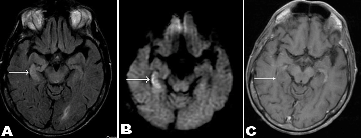
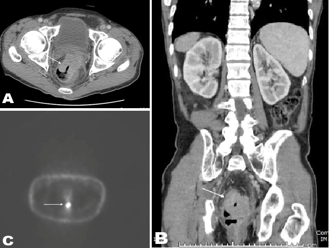
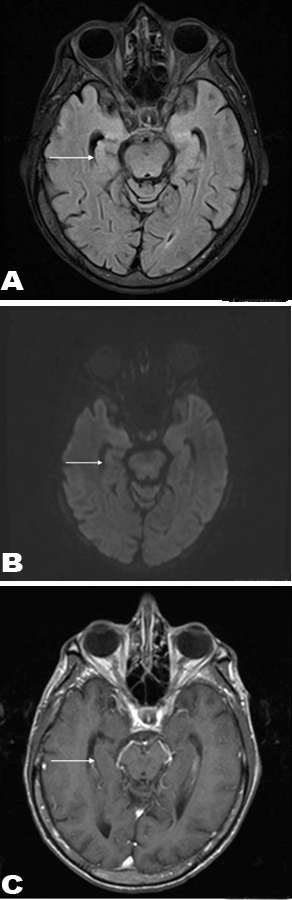
A 56-year-old male with recently diagnosed rectal cancer presented in the emergency department with recurrent seizures and disorientation for some days. Magnetic resonance imaging scan of brain demonstrated hyperintense signal on T2- FLAIR image (Figure 1A) and restricted diffusion on diffusion weighted image (Figure 1B) in right medial temporal lobe. There was no enhancement after contrast administration (Figure 1C). Patient's initial computed tomography (CT) scan of the abdomen and pelvis (Figure 2A-B), and positron emission tomography (PET) scan (Figure 2C) showed the presence of a large rectal mass with intense metabolic uptake. Based on clinical history and imaging findings, diagnosis of PLE was suggested. Serologic investigation showed a highly positive titer for anti-Ri (1:2560, normal <80). Subsequent cerebrospinal fluid analysis also showed increased titers of anti-Ri antibody with no findings to suggest viral or bacterial encephalitis. Since patient was already on chemotherapy treatment for rectal cancer, antiepileptic therapy and steroid treatment were also started. In next few days, the patient was free from seizure episodes and the neurological symptoms gradually improved. Follow-up MRI scan of brain was showed complete interval resolution of the abnormalities (Figure 3A-C). | ||||||
|
| ||||||
| ||||||
|
| ||||||
|
Discussion
| ||||||
|
Paraneoplastic neurologic syndromes are a group of neurologic disorders associated with malignancy affecting any part of the nervous system and include PLE, subacute cerebellar degeneration and sensory neuropathy [1]. Paraneoplastic limbic encephalitis one of the paraneoplastic neurologic syndromes, is caused by autoimmune-related inflammatory process localized to the limbic system. It is characterized by slowly progressive behavioral changes, memory loss, seizures and cognitive dysfunction [1]. Small cell lung cancer is most frequent associated malignancy. Other associated cancers include testicular tumors, breast cancer and Hodgkin lymphoma [2]. Infrequently, colon and rectal adenocarcinoma have also been shown to be associated with PLE. The most frequent antibodies are anti-Hu and anti-Ma2 antibodies which can be detected in serum and CSF, thus aiding in diagnosis [2]. Anti-Ri antibody is particularly associated with colon and rectal cancers [2]. As in any other intracranial abnormalities [3] [4], MRI scan is the imaging modality of choice. A brain MRI scan is helpful by demonstrating characteristic MRI finding of high signal intensity in bilateral or unilateral medial temporal lobes on T2-Flair images with mild restricted diffusion. Since DW images show restricted diffusion in the abnormal areas with cytotoxic edema and high cellularity [5] [6], PLE will also show restricted diffusion in most of the cases [7]. MRI scan also helps in excluding other pathologies with similar neurologic symptoms [8]. Differential diagnosis of MRI findings includes herpes encephalitis and low grade glioma. However, herpes encephalitis will usually involve the entire temporal lobe with involvement of the frontal lobe, with presence of hemorrhage in some cases. Low grade gliomas will be unilateral and usually not limited to medial temporal lobe [9]. In difficult cases, history of primary malignancy, serum and cerebrospinal fluid analysis help in making the correct diagnosis. Idiopathic limbic encephalitis can have similar MRI findings as PLE. However, they can usually be differentiated by the presence of high levels of voltage gated potassium channel (VGKC) antibodies serum and cerebrospinal fluid in cases of idiopathic limbic encephalitis [10]. But due to presence of antibodies in some patients with a paraneoplastic origin, the test cannot always be used to exclude a paraneoplastic cause of the limbic encephalitis. Thus, in these cases, overall clinical picture has to be taken into account before reaching the final diagnosis. Graus et al. suggested a diagnostic criterion for PLE which includes: a compatible clinical presentation; <4 years interval between tumor diagnosis and development of neurologic symptoms; exclusion of the other neurological complications of tumor and its treatment; and at least one of the following: MRI demonstrating temporal lobe abnormalities, cerebrospinal fluid with inflammatory changes but negative cytology and PCR for herpes encephalitis, or epileptic activity in the temporal lobe on EEG [11]. EEG findings in PLE include bilateral frontal and temporal intermittent rhythmic delta activity with disorganization and slowing of background activity [12]. Unlike other paraneoplastic syndromes of the central nervous system, PLE is well- known for its good response to therapy. Immunosuppression with methylprednisolone, intravenous immunoglobulin, and plasma exchange has been shown to be useful to control symptoms by reducing the titer of antibodies. However, treatment of associated tumor is most important in neurologic improvement and prognosis [13]. | ||||||
|
Conclusion
| ||||||
|
Paraneoplastic limbic encephalitis should be considered in the differential diagnosis of a patient having a history of malignancy and presenting with neurological symptoms. Magnetic resonance imaging helps in making the diagnosis by demonstrating signal abnormalities in the medial temporal lobes, and by excluding other causes of patient symptoms. Treatment of patient's primary malignancy and control of seizures with the help of antiepileptic drugs is the cornerstone of management of this condition, however immunosuppression with methylprednisolone, intravenous immunoglobulin, and plasma exchange is also shown to be useful to control symptoms by reducing the titer of antibodies. | ||||||
|
References
| ||||||
| ||||||
|
[HTML Abstract]
[PDF Full Text]
|
|
Author Contributions
Kusum Hooda – Substantial contributions to conception and design, Acquisition of data, Analysis and interpretation of data, Drafting the article, Revising it critically for important intellectual content, Final approval of the version to be published Nishant Gupta – Analysis and interpretation of data, Revising it critically for important intellectual content, Final approval of the version to be published Charu Chanana – Analysis and interpretation of data, Revising it critically for important intellectual content, Final approval of the version to be published Pranav Sharma – Analysis and interpretation of data, Revising it critically for important intellectual content, Final approval of the version to be published Salil Sharma – Analysis and interpretation of data, Revising it critically for important intellectual content, Final approval of the version to be published Joshua Sapire – Analysis and interpretation of data, Revising it critically for important intellectual content, Final approval of the version to be published |
|
Guarantor of submission
The corresponding author is the guarantor of submission. |
|
Source of support
None |
|
Conflict of interest
Authors declare no conflict of interest. |
|
Copyright
© 2017 Kusum Hooda et al. This article is distributed under the terms of Creative Commons Attribution License which permits unrestricted use, distribution and reproduction in any medium provided the original author(s) and original publisher are properly credited. Please see the copyright policy on the journal website for more information. |
|
|