| Table of Contents | |
|
Clinical Image
| ||||||
| Myeloid sarcoma of the breast soft tissue following allogeneic bone marrow transplantation | ||||||
| Jia-Ling Chou1, Lester D. R. Thompson1 | ||||||
|
1Southern California Permanente Medical Group, Department of Pathology, 5601 De Soto Avenue, Woodland Hills, CA.
| ||||||
| ||||||
|
[HTML Abstract]
[PDF Full Text]
[Print This Article]
[Similar article in Pumed] [Similar article in Google Scholar] |
| How to cite this article |
| Jia-Ling C, Thompson LDR. Myeloid sarcoma of the breast soft tissue following allogeneic bone marrow transplantation. J Case Rep Images Oncology 2016;2:71–73. |
|
Case Report
| ||||||
|
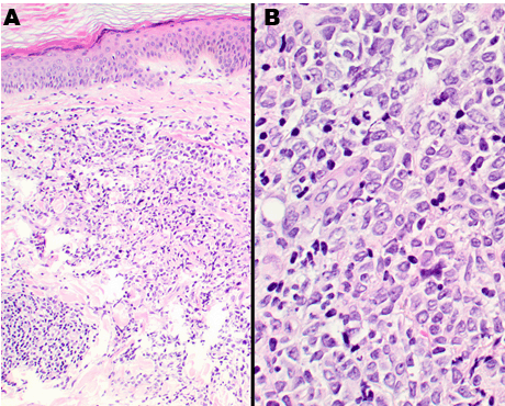
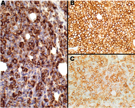
A 47-year-old female presented with high-risk myelodysplastic syndrome (MDS) without detectable cytogenetic abnormality from the bone marrow specimen. Initially, she received azacytidine while awaiting bone marrow transplantation (BMT). After successful allogeneic BMT from a mismatched related donor, she received post-transplant care including management of transplant-related toxicity and skin manifestations of graft versus host disease (GVHD) in a designated BMT center. Subsequent bone marrow showed no evidence of MDS, and she is currently free from GVHD. Approximately 1.5 years after BMT, the patient detected a palpable nodule in her right breast, just below the skin. Mammogram was negative. The lesion slowly enlarged over the next eight months, associated with brown skin discoloration. Breast ultrasound detected a 9-mm superficial mass with mixed echogenicity. Around the same time she found two hard lumps with similar skin color changes on the left and one on the right thighs. The lesions from the right breast and left thigh soft tissues were completely excised, while the right thigh mass was not excised, to be used as an index lesion to observe the effect of systemic therapy. The hematoxylin and eosin stained sections demonstrated infiltration of the dermis and subcutaneous soft tissues by aggregates of medium to large cells that had finely dispersed chromatin (Figure 1A-B). These cells stained strongly positive for myeloperoxidase (Figure 2A), CD 43 (Figure 2B), CD68 and CD117 (Figure 2C). The neoplastic cells were negative for CD3, CD5, CD20, CD34, CD10, CD30, ALK1, CD138, kappa, lambda, CK7 and pan-cytokeratin. The morphology and immunohistochemistry findings support the diagnosis of myeloid sarcoma (also known as granulocytic sarcoma or chloroma). A subsequent bone marrow aspiration and biopsy did not reveal leukemic cells and cytogenetic studies were completely normal. PET scan did not detect overly hypermetabolic activity apart from subcutaneous soft tissue lesions in the left medial thigh with SUV of 2.2, corresponding to one of the excised lesions. The patient is currently undergoing chemotherapy intended for acute myeloid leukemia (AML). | ||||||
|
| ||||||
|
| ||||||
| ||||||
|
Discussion
| ||||||
|
Myeloid sarcoma is formed by immature myeloid cells and is considered a rare extramedullary manifestation of acute myeloid leukemia (AML). It can occur concurrently with, following or, rarely, preceding AML. It can also be seen in MDS or chronic myeloid leukemia. It was reported among 0.2–1.3% of patients following BMT [1]. The symptoms of myeloid sarcoma may vary dependent on its location, rate of growth and size. It is frequently misdiagnosed as lymphoma [2] or Ewing's sarcoma. The most common sites of myeloid sarcoma include subcutaneous soft tissue, bone, lymph nodes, and periosteum. Myeloid sarcoma in breast soft tissue is rare [3] with only isolated case reports following BMT. It is reported that MS may precede AML by 5–12 months [4]. Therefore, treatment using the same regime for AML is called for once the diagnosis of myeloid sarcoma is established. | ||||||
|
Conclusion
| ||||||
|
As myeloid sarcoma in the breast soft tissue is very rare, it should be included in the differential diagnoses when a "breast lump" is found in a patient with non-lymphocytic hematological malignancy. Keywords: Acute myeloid leukemia, Breast, Immunohistochemistry, Myeloid sarcoma | ||||||
|
References
| ||||||
| ||||||
|
[HTML Abstract]
[PDF Full Text]
|
|
Author Contributions
Jia-Ling Chou – Substantial contribution to conception and design, Acquisition of data, Analysis and interpretation of the data, Drafting the article, Revising it critically for important intellectual content, Final approval of the version to be published Lester Thompson – Acquisition of data, Analysis and interpretation of the data, Revising it and critically evaluating manuscript for scientific content, Final approval of the version to be published |
|
Guarantor of submission
The corresponding author is the guarantor of submission. |
|
Source of support
None |
|
Conflict of interest
Authors declare no conflict of interest. |
|
Copyright
© 2016 Jia-Ling Chou et al. This article is distributed under the terms of Creative Commons Attribution License which permits unrestricted use, distribution and reproduction in any medium provided the original author(s) and original publisher are properly credited. Please see the copyright policy on the journal website for more information. |
|
|
|
About The Authors
| |||
| |||
| |||