| Table of Contents | |
|
Case Report
| ||||||
| Rapid growth of massive retinal gliosis in an infant | ||||||
| Nobuhiko Shiraki1, Takeshi Morimoto1, Takashi Fujikado1, Kohji Nishida1 | ||||||
|
1Department of Ophthalmology, Osaka University Graduate School of Medicine.
| ||||||
| ||||||
|
[HTML Abstract]
[PDF Full Text]
[Print This Article]
[Similar article in Pumed] [Similar article in Google Scholar] |
| How to cite this article: |
| Shiraki N, Morimoto T, Fujikado T, Nishida K. Rapid growth of massive retinal gliosis in an infant. J Case Rep Images Oncology 2015;1:28–31. |
|
Abstract
|
|
Introduction:
Massive retinal gliosis (MRG) is a rare, benign intraocular tumor that results from the proliferation of well-differentiated glial cells. MRGs develop because of a variety of causes.
Case Report: We encountered a case of a 2-year-old male infant with loss of vision in the right eye. Enucleation was carried out due to an absolute glaucoma. Histologically, the vitreous body had been totally replaced by massively proliferated spindle cells, which had delicate fibrillary cytoplasm without nuclear atypia. Immunohistochemically, the cells were strongly positive for glial fibrillary acidic protein. These findings led to a diagnosis of massive gliosis. Conclusion: Massive retinal gliosis (MRG) is a rare, benign intraocular tumor that results from the proliferation of well-differentiated glial cells. To our knowledge, this may be the first report of such an occurrence in a 2-year- old infant. It is important for ophthalmologists to be suspicious of a mass resistant to chemotherapy that resembles a RB in children. | |
|
Keywords:
Glavcoma, Infant tumor, Intraocular tumor, Massive retinal gliosis
| |
|
Introduction
|
|
A massive retinal gliosis (MRG) is a rare, benign intraocular tumor resulting from the proliferation of well-differentiated glial cells [1]. MRGs develop because of a variety of causes [2]. We present a case of MRG in a young male child. |
|
Case Report
|
|
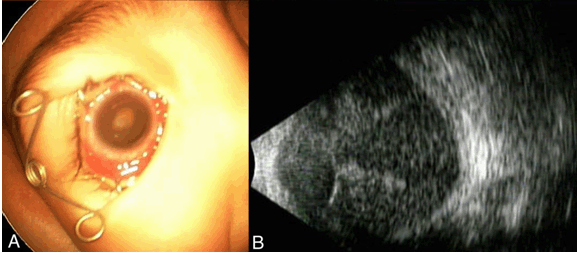
A healthy two-year-old boy presented with leukocoria of the right eye. On examination, the boy had poor visual fixation in the right eye and steady fixation in his left eye. His left eye decimal visual acuity was 0.4, and the right eye was light perception. Indirect ophthalmoscopy revealed a white tumor which resembled a retinoblastoma (Figure 1A). Ultrasonography showed a total retinal detachment and a lesion in the subretinal space with no calcification (Figure 1B). Computed tomography (CT) scan of head showed that the tumor was located in vitreous cavity of the right eye without calcification (Figure 1C). Whole body CT scan revealed no other tumors. We suspected a retinoblastoma and performed 2 cycles of chemotherapy consisting of vincristine, etoposide, and carboplatin (VEC). However, the VEC treatment had no effect, and the tumor grew rapidly (Figure 2A-B). The intraocular pressure (IOP) of the right eye increased to >60 mmHg. Finally, the vision in his right eye decreased to no light perception (NLP). The patient also lost his appetite due to the severe pain of right eye. Given the uncertainty over the clinical diagnosis, the poor visual prognosis, sever pain, and absolute glaucoma, the right eye was enucleated. His state was improved after the operation. Histopathologic studies showed an endophytic mass in the vitreous cavity with vitreous hemorrhage. The tumor was composed of spindle-shaped, well differentiated cells with small nuclei and abundant, pale, eosinophilic cytoplasm especially in the inner retinal layers (Figure 3A-B). For immunohistochemical studies, 4-µm-thick sections were stained with GFAP (dilution at x200 without pretreatment, DakoCytomation, Kyoto, Japan), anti-NSE (no dilution without pretreatment, Nichirei, Tokyo, Japan), anti-S-100 protein (no dilution and without pretreatment, Nichirei), and anti-Ki-67 antibodies (MIB-1, x50 after pretreatment by autoclaving for 15 minutes, DakoCytomation) [3]. The cytoplasm of the proliferating spindle cells was diffusely positive for GFAP (Figure 3C), but NSE and S-100 protein staining were not detected (Figure 3D, E). The cells in the inner retinal layer were more immunopositive for Ki-67 (Figure 3F). These findings led to a diagnosis of massive retinal gliosis. |
|
|
|
|
|
|
|
Discussion
|
|
There have been several reports of MRG of the orbit [1] [2] [3] [4] [5] [6] [7] [8] [9] [10], but our case was unique because it developed in an infant. This tumor was also unique because its funduscopic appearance strongly resembled the typical findings of retinoblastomas by ultrasonography. In addition, the CT scan of head showed a tumor without calcification. Other possible intraocular tumors include choroidal melanoma, astrocytic hamartoma, retinal hemangioblastomas, tumors of retinal pigment epithelium, intraocular metastasis, and vasoproliferative tumors of the retina [4] [5] [6] [7]. Therefore, it was difficult to diagnose MRG based on our clinical finding without histopathological examinations. Massive glial proliferations occur in eyes with underlying pathologic processes such as retinitis pigmentosa [8], retinopathy of prematurity [9], longstanding retinal detachment [6], disc abnormality [10], central retinal vein occlusion, or trauma [2]. However, proliferation can occur without a history of ocular or other diseases [11]. In our patient, there was no signs suggesting any of these tumors. Yanoff et al. reviewed 38 histologically diagnosed MRG cases and reported that both sexes and all ages may be affected with nearly equal frequency [2]. However, the age of the patients ranged 20 to 70 years in other reports [4] [5] [6] [8] [9] [10] [11]. Histologically, MRG has been shown to be non-neoplastic and originate from Müller cells [1] [3][11]. Histological findings showed that the retina can be totally replaced by proliferated uniform spindle cells with abundant, eosinophilic, fibrillated cytoplasm and indistinct cell borders. Bruch's membrane was intact and the vitreous cavity was filled with the same spindle cells in some patients [1] [3][9][11]. Immunohistochemistry showed that the spindle and oval cells were positive to GFAP and neuron specific enolase (NSE) and partly with S-100 protein [3] [11]. The histology and immunohistochemical characteristic of our patient were similar to that reported. In our case, the rapid growth of the mass suggested malignancy, and RB appeared to be the most likely diagnosis. However, chemotherapy was ineffective, and the eye was eventually enucleated. Only after histological and immunohistochemical studies were we able to diagnose the mass as MRG. So, it is important for ophthalmologists to be suspicious of a mass that resembles a ratinoblastoma in children. If chemotherapy has no effect, then enucleation should be considered when the eye is blind and painful. |
|
Conclusion
|
|
Massive retinal gliosis (MRG) is a rare, benign intraocular tumor that results from the proliferation of well-differentiated glial cells. To our knowledge, this may be the first report of such an occurrence in a two-year-old boy. It is important for ophthalmologists to be suspicious of a mass resistant to chemotherapy that resembles a ratinoblastoma in children. |
|
Acknowledgements
|
|
This study was conducted without any outside commer-cial interests and has not been previously published. |
|
References
|
|
|
[HTML Abstract]
[PDF Full Text]
|
|
Author Contributions
Nobuhiko Shiraki – Substantial contributions to conception and design, Acquisition of data, Analysis and interpretation of data, Drafting the article, Revising it critically for important intellectual content, Final approval of the version to be published Takeshi Morimoto – Analysis and interpretation of data, Revising it critically for important intellectual content, Final approval of the version to be published Takashi Fujikado – Analysis and interpretation of data, Revising it critically for important intellectual content, Final approval of the version to be published Kohji Nishida – Analysis and interpretation of data, Revising it critically for important intellectual content, Final approval of the version to be published |
|
Guarantor of submission
The corresponding author is the guarantor of submission. |
|
Source of support
None |
|
Conflict of interest
Authors declare no conflict of interest. |
|
Copyright
© 2015 Nobuhiko Shiraki et al. This article is distributed under the terms of Creative Commons Attribution License which permits unrestricted use, distribution and reproduction in any medium provided the original author(s) and original publisher are properly credited. Please see the copyright policy on the journal website for more information. |
|
|